. 2014 Feb 10;25(2):210-25.
doi: 10.1016/j.ccr.2014.01.028.

Disrupting the interaction of BRD4 with diacetylated Twist suppresses tumorigenesis in basal-like breast cancer

Affiliations

Disrupting the interaction of BRD4 with diacetylated Twist suppresses tumorigenesis in basal-like breast cancer

Jian Shi et al. Cancer Cell. .

Abstract

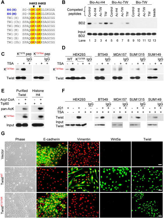
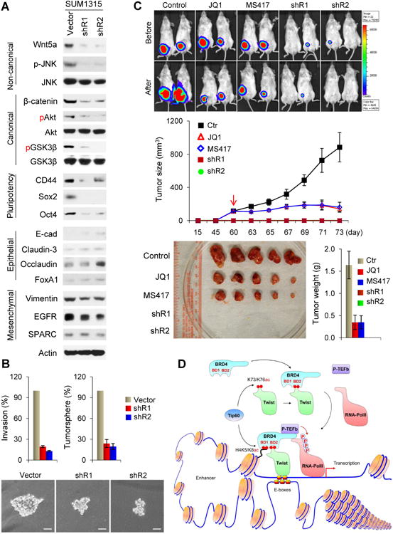
Twist is a key transcription activator of epithelial-mesenchymal transition (EMT). It remains unclear how Twist induces gene expression. Here we report a mechanism by which Twist recruits BRD4 to direct WNT5A expression in basal-like breast cancer (BLBC). Twist contains a "histone H4-mimic" GK-X-GK motif that is diacetylated by Tip60. The diacetylated Twist binds the second bromodomain of BRD4, whose first bromodomain interacts with acetylated H4, thereby constructing an activated Twist/BRD4/P-TEFb/RNA-Pol II complex at the WNT5A promoter and enhancer. Pharmacologic inhibition of the Twist-BRD4 association reduced WNT5A expression and suppressed invasion, cancer stem cell (CSC)-like properties, and tumorigenicity of BLBC cells. Our study indicates that the interaction with BRD4 is critical for the oncogenic function of Twist in BLBC.


Conflict of interest statement

Conflict of interest statement: The authors have declared that no conflict of interest exists

Figures

Figure 1
Figure 1. BD2 of BRD4 is required for its interaction with acetylated Twist
(A) HA-Twist and Flag-BRD4 were co-expressed in HEK293 cells. After treating cells with TSA (2 μM) for 12 hr, Twist, BRD4 and acetylated Twist were immunoprecipitated with HA, Flag and pan-acetylated-lysine (pan-AcK) antibodies, respectively, and analyzed by Western blotting. (B) HeLa cells stably expressing Flag-Twist were treated with TSA as in (A), Flag-Twist, endogenous BRD4 and acetylated Twist were immunoprecipitated and examined by Western blotting. (C) Cells were treated as described in (A), endogenous Twist, BRD4 and acetylated Twist were immunoprecipitated and examined by Western blotting. (D) Schematic depiction of the functional domains of BRD4 and deletion constructs used (top panel). ET stands for extra-terminal domain. Flag-tagged wild-type (WT) or deletion mutants of BRD4 were co-expressed with HA-Twist in HEK293 cells. After immunoprecipitated with HA or Flag antibody, the bound BRD4 or Twist was examined by Western blotting. (E) Schematic diagram showing the double bromodomain (BD1+BD2) of BRD4 and individual BD constructs used (top panel). Flag-BD1WT, BD1YN, BD2WT and BD2YN were co-expressed with HA-Twist in HEK293 cells treated with TSA as in (A). Twist and BDs were immunoprecipitated with HA and Flag antibodies, respectively, and analyzed by Western blotting. (F) HA-Twist and Flag-BRD4 were co-expressed in HEK293 cells treated with TSA as in (A). Twist and BRD4 were immunoprecipitated with HA and Flag antibodies, respectively, in the presence or absence of JQ1 (1 μM) and analyzed by Western blotting. (G) Cells were treated with TSA as in (A), endogenous Twist and BRD4 were immunoprecipitated with Twist and BRD4 antibodies, respectively, in the presence or absence of JQ1 (1 μM) and examined by Western blotting. See also Figure S1
Figure 2
Figure 2. Twist di-acetylation at K73/K76 by Tip60 is required for interaction with BRD4
(A) Schematic diagram showing the domain organization of Twist, with deletion and mutation constructs used (top panel). HA-tagged WT or deletion mutants of Twist were co-expressed with Flag-BRD4 in HEK293 cells treated with or without TSA. Twist and BRD4 were immunoprecipitated with HA and Flag antibodies, respectively, and analyzed by Western blotting. (B) HA-tagged WT or deletion mutants of Twist were expressed in HEK293 cells treated with TSA. Twist and acetylated Twist were immunoprecipitated with HA and pan-AcK antibodies, respectively, and analyzed by Western blotting. (C) Flag-tagged WT or deletion mutants of Twist were co-expressed with HA-Tip60 in HEK293 cells treated with TSA. Twist and Tip60 were immunoprecipitated with Flag and HA antibodies, respectively, and analyzed by Western blotting. (D) Ectopic expression of Tip60 or knockdown of endogenous Tip60 was performed in BT549 and SUM1315 cells. After endogenous Twist was immunoprecipitated, acetylation of Twist and the bound BRD4 were examined by Western blotting. (E) HA-tagged WT and mutant Twist were expressed in HEK293 cells, acetylated Twist was immunoprecipitated with HA and pan-AcK antibodies, respectively, and examined by Western blotting. (F) HA-tagged WT or mutant Twist was co-expressed with Flag-tagged BD1 and BD2 in HEK293 cells treated with TSA. Twist and BDs were immunoprecipitated with HA and Flag antibodies, respectively, and examined by Western blotting. (G) HA-tagged WT or mutant Twist was co-expressed with Flag-tagged BD2 in HEK293 cells treated with TSA. Twist, BRD4, and acetylated Twist were immunoprecipitated with HA, Flag, and pan-AcK antibodies, respectively, and analyzed by Western blotting. (H) Determination of Tip60 catalyzed acetylation sites in Twist by mass spectrometry. Peptides contain K73 and K76 acetylations are shown in the top and bottom panels, respectively. See also Figure S2
Figure 3
Figure 3. K73ac/K76ac Twist and BRD4 interaction is critical for the function of Twist
(A) Sequence alignment between K5/8 of histone H4 and K73/76 of Twist. H, M, Ch, Mk, R, B, and Sh stand for human, mouse, chimpanzee, monkey, rat, bovine, and sheep, respectively. (B) The indicated biotinylated peptides were mixed with lysates from HEK293 cells expressing BD2 without or with indicated non-biotinylated competing peptides, the bound BD2 was analyzed by Western blotting after pull-down of the biotinylated peptides. (C) HA-Twist was expressed in HEK293 cells treated with or without TSA. The immunoprecipitated Twist was analyzed on Western blots using indicated antibody in the presence of Twist-K73/K76 or Twist-K73ac/K76ac peptides. (D) HA-tagged or endogenous Twist was immunoprecipitated from cells treated with or without TSA using HA and Twist antibodies, respectively, and analyzed by K73ac/K76ac antibody. (E) Purified human Twist or histone H4 was incubated with purified Tip60 in the absence or presence of acetyl-CoA. The acetylation of Twist and histone H4 was examined by pan-AcK and anti-K73ac/K76ac antibodies. (F) K73ac/K76ac Twist was immunoprecipitated with K73ac/K76ac antibody in the presence or absence of JQ1. The bound Twist was examined by Western blotting. (G) HMLE cells expressing the vector or the WT or K73/76R Twist were examined for morphological changes indicative of EMT by phase-microscopy and the expression of E-cadherin, vimentin, Wnt5a, and Twist (green) by immunofluorescent staining. Nuclei stained with DAPI (red). Scale bar, 50 μM. See also Figure S3
Figure 4
Figure 4. The structural and molecular basis of Twist-K73ac/K76ac recognition by BRD4
(A) Stereo ribbon diagram of the 3D solution structure of the BRD4-BD2 bound to a di-acetylated K73ac/K76ac Twist peptide (yellow). Side chains of key residues engaged at the protein/peptide interactions are depicted and color-coded by atom type. (B) Surface electrostatic potential (left) or Space-filled (right) representation of the BRD4-BD2/Twsit-K73ac/K76ac complex structure highlights His437 (red) at the acetyl-lysine binding site that is responsible for the BRD4-BD2' specificity of this molecular recognition. (C) HA-tagged Twist was co-expressed with Flag-tagged WT or mutant BD1 and BD2 in HEK293 cells. Twist and BDs were immunoprecipitated with HA and Flag antibodies, respectively, and the bound BDs and Twist were analyzed by Western blotting. (D) HA-tagged Twist was co-expressed with Flag-tagged BD1, BD2, BD1+BD2 in HEK293 cells. After immunoprecipitated Twist, BDs and H4, the association and acetylation of these molecules were examined by Western blotting. See also Figure S4 and Table S1
Figure 5
Figure 5. Twist positively correlates with Wnt5a expression in breast cancer
(A) Gene expression profiling analysis (left panel) was used to identify potential Twist target genes. Common Twist target genes between HMLE and T47D cells were shown in the heat-map (right panel). (B) The mRNA and protein levels of Twist, Wnt5a and BRD4 were analyzed by RT-PCR and Western blotting. (C) The Effects of stable shRNA knockdown of endogenous Twist in SUM1315 cells that were transiently transfected with WT or mutant (KR) Twist on the expression of Wnt5a and various molecules was evaluated by Western blotting. (D) Wnt5a expression in five BLBC cell lines with knockdown of Twist and/or BRD4 was analyzed by Western blotting. NTC stands for non-target control siRNA. See also Figure S5
Figure 6
Figure 6. The Twist-BRD4 complex directly activates WNT5A transcription
(A) Schematic depiction of the WNT5A promoter and WNT5A reporter luciferase constructs used (top panel). Enhancement of Wnt5a luciferase activity by co-expression of Twist and BRD4 in HEK293 cells. (B) Twist, BRD4, H4ac4, RNA-PolII and P-TEFb (CDK9) association at the WNT5A promoter as assessed by ChIP. SP-specific primer; CP-control primer (5 kb downstream of 3′-UTR). (C) Twist, BRD4, H4ac and H3K27ac association at the WNT5A enhancer as assessed by ChIP SP-specific primer (22 kb upstream of TSS); CP-control primer (5 kb downstream of 3′-UTR). (D) Effects of Twist-knockdown or JQ1 treatment on the association of Twist, BRD4, RNA-PolII and P-TEFb (CDK9) and H4ac4 at the WNT5A promoter as assessed by ChIP in SUM1315 cells. SP and CP primers used are same as in (B). (E) (Left panel) Assessing effects of transient expression of HA-Twist and/or Flag-BRD4 on their association with RNA-PolII and P-TEFb (CDK9) in HEK293 cells. (Right panel) Assessment of endogenous BRD4 knockdown effect on the association of Twist, BRD4, RNA-PolII and P-TEFb (CDK9) in SUM1315 cells. (F) SUM1315 cells were serum starved for overnight followed by stimulation with FGF, TNFα, or EGF plus insulin for 3 hr in the absence or presence of JQ1 (right panel). Twist was immunoprecipitated and K73ac/K76ac of Twist and the association of Tip60 and BRD4 were analyzed. Expression of Wnt5a, Twist, BRD4 and Tip60 were also examined by Western blotting. (G) BLBC cells were treated with JQ1, expression of c-Myc and Wnt5a was analyzed by Western blotting. (H) Invasion (left) and tumorsphere-formation assays (right) of cells treated as in (G) were examined in the absence or present of recombinant Wnt5a (100 ng/ml). Statistical analysis (mean ± SD) from three independent experiments with duplicates was shown. For A, B, C, and D, statistical analysis (mean ± SD) from three separate experiments in triplicates was shown. See also Figure S6
Figure 7
Figure 7. The Twist-BRD4-Wnt5a axis is critical for tumorigenicity in vitro and in vivo
(A) Expression of various molecules in SUM1315 cells with Wnt5a-knockdown. (B) Invasion and tumorsphere-formation in SUM1315 cells with Wnt5a-knockdown. Data are presented as a percentage of vector control values (mean ± SD in three separate experiments in duplicates). Representative pictures of tumorsphere are shown in the bottom panel, Scale bar, 100 μM. (C) Vector control and Wnt5a-knockdown SUM1315 cells were injected into the mammary fat pad of NOD-SCID mice. When tumors from mice injected with control cells reached 100 mm3, mice were divided into three groups and treated with JQ1 (50 mg/Kg), MS417 (20 mg/Kg), or solvent control, respectively. The size of tumor was recorded by bioluminescence imaging before or after 2-wk treatment. Tumor weight was also measured. Data are represented as a mean ± SEM from 5 mice. (D) A proposed model illustrating the interaction of Twist and BRD4 at the enhancer/promoter of WNT5A, which leads to the transcriptional activation of WNT5A expression in EMT and BLBC. See also Figure S7

References

    1. Brunger AT. X-PLOR version 3.1: A system for X-ray Crystallography and NMR (version 3.1 edit) Yale University Press; New Haven, CT: 1993.
    1. Canzio D, Liao M, Naber N, Pate E, Larson A, Wu S, Marina DB, Garcia JF, Madhani HD, Cooke R, et al. A conformational switch in HP1 releases auto-inhibition to drive heterochromatin assembly. Nature. 2013;496:377–381. - PMC - PubMed
    1. Clore GM, Gronenborn AM. Multidimensional heteronuclear nuclear magnetic resonance of proteins. Methods in enzymology. 1994;239:349–363. - PubMed
    1. Dawson MA, Prinjha RK, Dittmann A, Giotopoulos G, Bantscheff M, Chan WI, Robson SC, Chung CW, Hopf C, Savitski MM, et al. Inhibition of BET recruitment to chromatin as an effective treatment for MLL-fusion leukaemia. Nature. 2011;478:529–533. - PMC - PubMed
    1. Delmore JE, Issa GC, Lemieux ME, Rahl PB, Shi J, Jacobs HM, Kastritis E, Gilpatrick T, Paranal RM, Qi J, et al. BET bromodomain inhibition as a therapeutic strategy to target c-Myc. Cell. 2011;146:904–917. - PMC - PubMed

Publication types

MeSH terms

Associated data