

doi: 10.1038/ncomms4309.
Lipidic cubic phase injector facilitates membrane protein serial femtosecond crystallography
Affiliations
- PMID: 24525480
- PMCID: PMC4061911
- DOI: 10.1038/ncomms4309
Item in Clipboard
Lipidic cubic phase injector facilitates membrane protein serial femtosecond crystallography
Nat Commun.
2014.
Abstract
Lipidic cubic phase (LCP) crystallization has proven successful for high-resolution structure determination of challenging membrane proteins. Here we present a technique for extruding gel-like LCP with embedded membrane protein microcrystals, providing a continuously renewed source of material for serial femtosecond crystallography. Data collected from sub-10-μm-sized crystals produced with less than 0.5 mg of purified protein yield structural insights regarding cyclopamine binding to the Smoothened receptor.
Conflict of interest statement
The authors declare no competing financial interests.
Figures
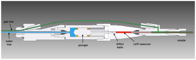
In operation, the device is attached via the leftmost threaded fitting to a nozzle rod (not shown) for insertion into the experimental chamber. Water (blue) and gas (green) lines are routed through the nozzle rod from the left, LCP (red) is extruded from the nozzle on the right. Water, at a pressure of up to 300 psi, drives the hydraulic plunger, which amplifies the pressure 34 times to drive LCP through a capillary with an inner diameter of 10–50 μm. Two spherical Teflon beads are used to provide a tight seal against a pressure of up to 10,000 psi. The co-flowing gas is necessary for reliable extrusion and to maintain co-axial flow.

Panels (a) and (b): Single femtosecond snapshot diffraction patterns. (a) Diffraction spots from A2A adenosine receptor microcrystals in 9.9 MAG/cholesterol LCP to 2.5 Å and strong powder diffraction rings from crystalline lipid. (X-ray intensity attenuated to 7%, 1.5 μm X-ray beam diameter, 50 fs pulse length, 9.5 keV, 15 μm LCP jet diameter, 300 pL/min flow rate, 1 Hz pulse rate, crystal size: 1×1×5 μm3). (b) Diffraction from serotonin receptor 5-HT2B in cholesterol-doped 9.9 MAG + 7.9 MAG LCP. No sharp rings are visible suggesting that formation of Lc phase has been avoided (X-ray intensity attenuated to 3.1% due to strong Bragg diffraction from 5×5×5 μm3 sized crystals, 1.5 μm X-ray beam diameter, 50 fs pulse length, 9.5 keV, 50 μm LCP jet diameter, 190 nL/min flow rate, 120 Hz pulse rate). The resolution at the detector edge in both panels is 2.5 Å. Panels (c) and (d): 9.9 MAG LCP extrusion in vacuum viewed between crossed polarizers. The tapered end of the capillary nozzle is seen protruding out of the gas aperture. Capillary inner diameter: 30 μm. (c) with He as co-flowing gas. Birefringence (bright flecks) is an indication of a transition of the cubic phase to a lamellar crystalline phase due to evaporative cooling. (d) with N2 as co-flowing gas and no visible birefringence. Scale bars: 100 μm.
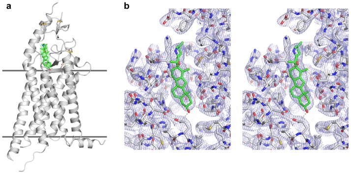
(a) Receptor model is shown as a gray cartoon, cyclopamine as a stick model with green carbons, and the “omit” 2mFo-DFc density map for cyclopamine contoured at 1 σ is shown as green wires. Horizontal lines indicate membrane boundaries. (b) Cyclopamine binding pocket is shown in stereo view as a stick model along with 2mFo-DFc density map contoured at 1 σ. Cyclopamine binds near to the entrance into a long and narrow cavity inside the receptor. Polar interactions stabilizing the shape of the cavity and cyclopamine binding are shown as gray dashed lines. Viewing angles in (a) and (b) are slightly different.
Comment in
-
Making protein crystals fly.Nat Methods. 2014 Apr;11(4):366-7. doi: 10.1038/nmeth.2913. Nat Methods. 2014. PMID: 24818225 No abstract available.
-
Serial femtosecond crystallography datasets from G protein-coupled receptors.Sci Data. 2016 Aug 1;3:160057. doi: 10.1038/sdata.2016.57. Sci Data. 2016. PMID: 27479354 Free PMC article.
References
-
- Yildirim MA, Goh KI, Cusick ME, Barabási AL, Vidal M. Drug-target network. Nat Biotechnol. 2007;25:1119–1126. - PubMed
-
- Deisenhofer J, Epp O, Miki K, Huber R, Michel H. Structure of the protein subunits in the photosynthetic reaction centre of Rhodopseudomonas viridis at 3 Å resolution. Nature. 1985;318:618–624. - PubMed
Publication types
MeSH terms
Substances
Associated data
- Actions
Grants and funding
LinkOut - more resources
Full Text Sources
Other Literature Sources
Molecular Biology Databases
