. 2014 Feb 13;5(2):e1056.
doi: 10.1038/cddis.2014.33.

Nonthermal plasma induces head and neck cancer cell death: the potential involvement of mitogen-activated protein kinase-dependent mitochondrial reactive oxygen species

Affiliations

Nonthermal plasma induces head and neck cancer cell death: the potential involvement of mitogen-activated protein kinase-dependent mitochondrial reactive oxygen species

S U Kang et al. Cell Death Dis. .

Abstract

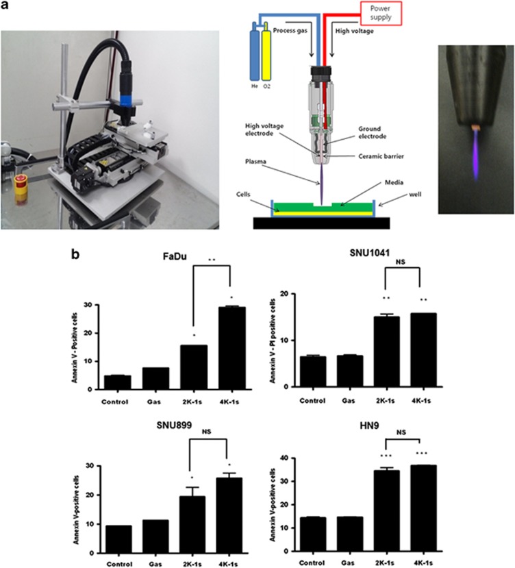
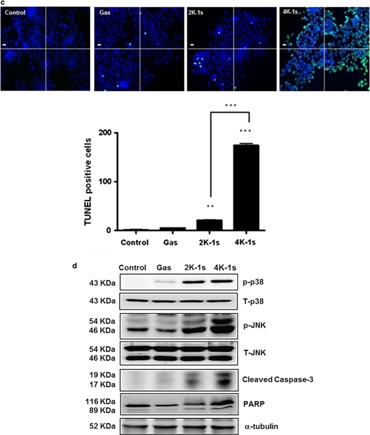
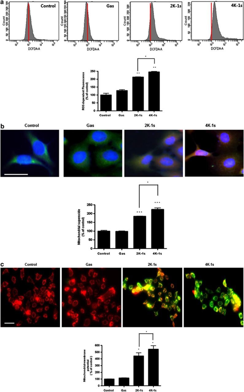
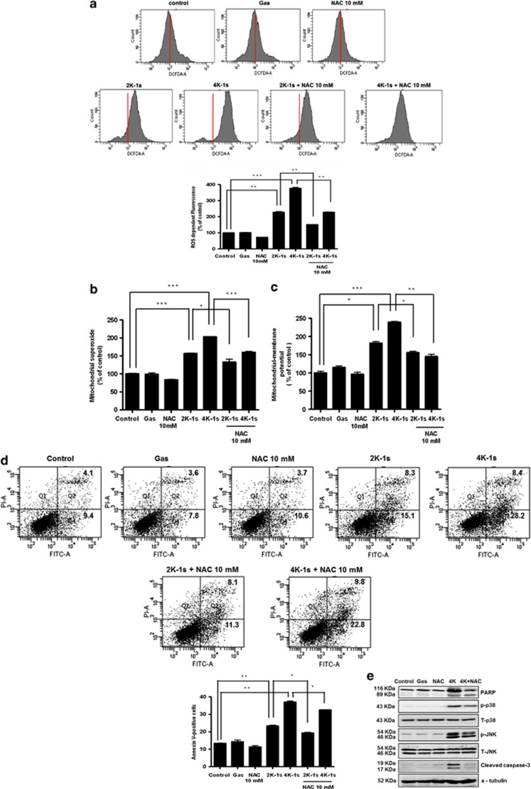
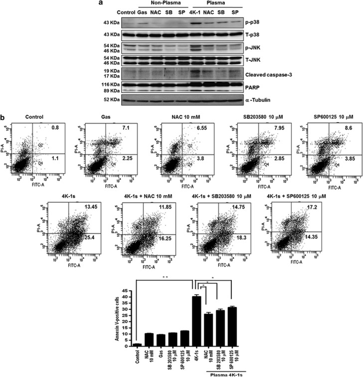
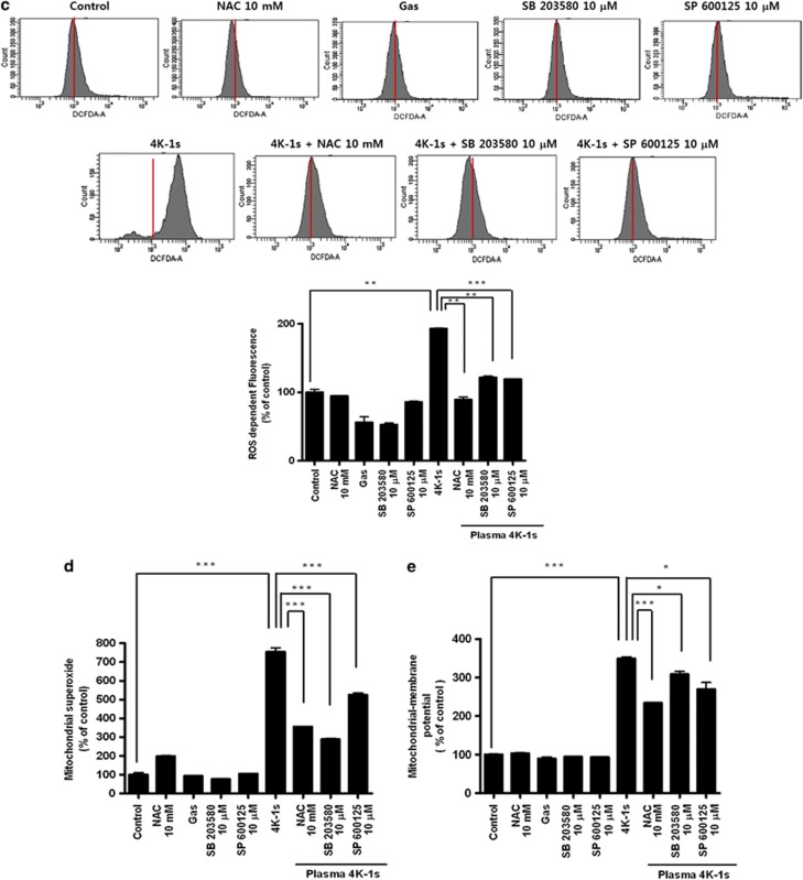
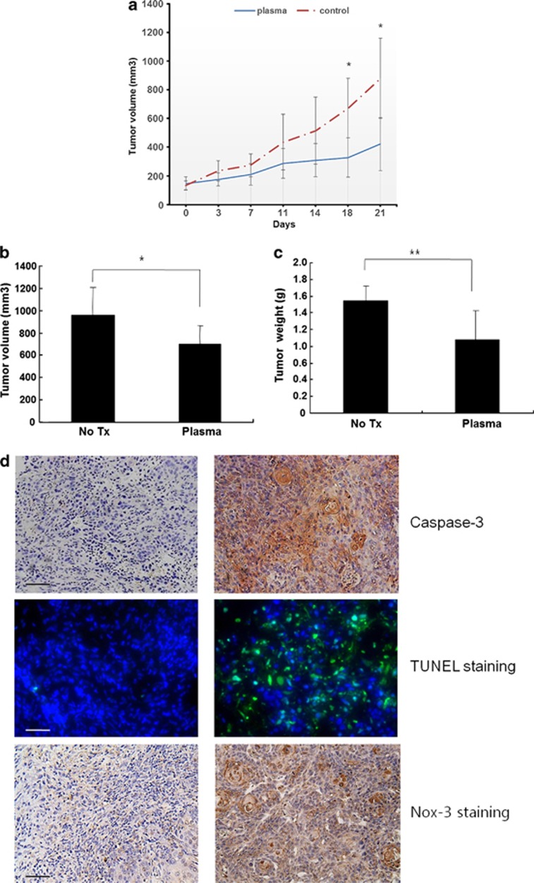
Nonthermal plasma (NTP) is generated by ionization of neutral gas molecules, which results in a mixture of energy particles including electrons and ions. Recent progress in the understanding of NTP has led to its application in the treatment of various diseases, including cancer. However, the molecular mechanisms of NTP-induced cell death are unclear. The purpose of this study was to evaluate the molecular mechanism of NTP in the induction of apoptosis of head and neck cancer (HNC) cells. The effects of NTP on apoptosis were investigated using MTT, terminal deoxynucleotidyl transferase-mediated dUTP-biotin nick-end labeling, Annexin V assays, and western blot analysis. The cells were examined for production of reactive oxygen species (ROS) using DCFCA or MitoSOX staining, intracellular signaling, and an animal model. NTP reduced HNC cell viability in a dose-dependent manner and induced apoptosis. NTP resulted in alteration of mitochondrial membrane potential and accumulation of intracellular ROS generated from the mitochondria in HNC cells. Blockade of ROS production by N-acetyl-L-cysteine inhibited NTP-induced apoptosis. NTP led to the phosphorylation of c-JUN N-terminal kinase (JNK) and p38, but not extracellular-regulated kinase. Treatment with JNK and p38 inhibitors alleviated NTP-induced apoptosis via ROS generation. Taken together, these results show that NTP induced apoptosis of HNC cells by a mechanism involving MAPK-dependent mitochondrial ROS. NTP inhibited the growth of pre-established FaDu tumors in a nude mouse xenograft model and resulted in accumulation of intracellular ROS. In conclusion, NTP induced apoptosis in HNC cells through a novel mechanism involving MAPK-mediated mitochondrial ROS. These findings show the therapeutic potential of NTP in HNC.


Figures

Figure 1
Figure 1
Apoptotic effects of NTP on HNC cells. (a) Photograph of a plasma jet, schematic diagram of the plasma system, and image of a plasma jet. (b) Various HNC cells were analyzed 24 h after treatment with NTP by staining with Annexin V/PI. (c) Apoptosis of FaDu cells was determined by the TUNEL method using a detection kit. (d) The cell lysates were then separated by SDS-PAGE and immunoblotted with anti-p-JNK, p-p38, p-ERK, cleaved caspase-3, and PARP antibodies. Scale bar denotes 50 μm. *P<0.05, **P<0.01, and ***P<0.001
Figure 1
Figure 1
Apoptotic effects of NTP on HNC cells. (a) Photograph of a plasma jet, schematic diagram of the plasma system, and image of a plasma jet. (b) Various HNC cells were analyzed 24 h after treatment with NTP by staining with Annexin V/PI. (c) Apoptosis of FaDu cells was determined by the TUNEL method using a detection kit. (d) The cell lysates were then separated by SDS-PAGE and immunoblotted with anti-p-JNK, p-p38, p-ERK, cleaved caspase-3, and PARP antibodies. Scale bar denotes 50 μm. *P<0.05, **P<0.01, and ***P<0.001
Figure 2
Figure 2
Induction of ROS in NTP-treated FaDu cells. (a) FaDu cells were treated with DCFDA and assayed using flow cytometry. (b) For measurement of mitochondrial superoxide, the cells were incubated with 2.5 μM of MitoSOX and then stained with 180 nM MitoTracker. (c) MMP was measured by flow cytometry using JC-1 fluorescence. Scale bar denotes 50 μm. *P<0.05, **P<0.01, and ***P<0.001
Figure 3
Figure 3
Effect of NTP-generated ROS on apoptosis. Cells were treated with NAC (10 mM) for 1 h before treatment with NTP. (a) Measurement of ROS generation using flow cytometry. (b) Measurement of mitochondrial superoxide with MitoSOX and MitoTracker. (c) Measurement of MMP with JC-1. (d) Analysis of apoptosis by FACS with Annexin V-PI. (e) The cell lysates were assessed by western blot analysis using antibodies against p-JNK, p-p38, p-ERK, cleaved caspase-3, and PARP. *P<0.05, **P<0.01, and ***P<0.001
Figure 4
Figure 4
Involvement of MAPK in the generation of mitochondrial ROS and mitochondrial dysfunction. Apoptosis of FaDu cells was assessed 24 h after NTP with or without preincubation with NAC (10 mM), SB2023580 (10 μM), or SP 600125 (10 μM). (a) Immunoblotting was performed using antibodies against p-JNK, p-p38, p-ERK, cleaved caspase-3, and PARP. (b) Analysis of apoptosis by FACS with Annexin V-PI. (c) Measurement of ROS generation by flow cytometry. (d) Measurement of mitochondrial superoxide using MitoSOX and MitoTracker. (e) Measurement of MMP using JC-1. *P<0.05, **P<0.01, and ***P<0.001
Figure 4
Figure 4
Involvement of MAPK in the generation of mitochondrial ROS and mitochondrial dysfunction. Apoptosis of FaDu cells was assessed 24 h after NTP with or without preincubation with NAC (10 mM), SB2023580 (10 μM), or SP 600125 (10 μM). (a) Immunoblotting was performed using antibodies against p-JNK, p-p38, p-ERK, cleaved caspase-3, and PARP. (b) Analysis of apoptosis by FACS with Annexin V-PI. (c) Measurement of ROS generation by flow cytometry. (d) Measurement of mitochondrial superoxide using MitoSOX and MitoTracker. (e) Measurement of MMP using JC-1. *P<0.05, **P<0.01, and ***P<0.001
Figure 5
Figure 5
Effect of NTP on tumor growth and induction of apoptosis in vivo. (a) Sixteen mice were randomly divided into two groups and treated with NTP daily for 20 s. Tumor volumes were measured using a caliper two times per week. (b) Tumor volume and (c) weight were measured after they were killed. (d) Caspase-3, Nox-3, and TUNEL assays were performed on the tissues excised from the mice that were on day 21. Scale bar denotes 200 μm. *P<0.05 and **P<0.01

References

    1. Jemal A, Siegel R, Ward E, Hao Y, Xu J, Thun MJ. Cancer statistics, 2009. Cancer J Clin. 2009;59:225–249. - PubMed
    1. Argiris A, Karamouzis MV, Raben D, Ferris RL. Head and neck cancer. Lancet. 2008;371:1695–1709. - PMC - PubMed
    1. Kim CH, Kwon S, Bahn JH, Lee K, Jun SI, Rack PD, et al. Effects of atmospheric nonthermal plasma on invasion of colorectal cancer cells. Appl Phys Lett. 2010;96:243701. - PMC - PubMed
    1. Sensenig R, Kalghatgi S, Cerchar E, Fridman G, Shereshevsky A, Torabi B, et al. Non-thermal plasma induces apoptosis in melanoma cells via production of intracellular reactive oxygen species. Ann Biomed Eng. 2011;39:674–687. - PMC - PubMed
    1. Kim CH, Bahn JH, Lee SH, Kim GY, Jun SI, Lee K, et al. Induction of cell growth arrest by atmospheric non-thermal plasma in colorectal cancer cells. J Biotechnol. 2010;150:530–538. - PubMed

Publication types

MeSH terms