. 2014 Apr 1;74(7):2082-93.
doi: 10.1158/0008-5472.CAN-12-0452. Epub 2014 Feb 13.

Small GTPase RhoE/Rnd3 is a critical regulator of Notch1 signaling

Affiliations

Small GTPase RhoE/Rnd3 is a critical regulator of Notch1 signaling

Zehua Zhu et al. Cancer Res. .

Abstract

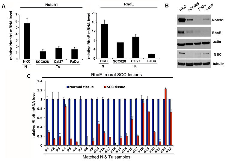
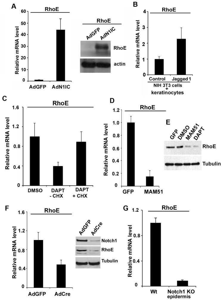
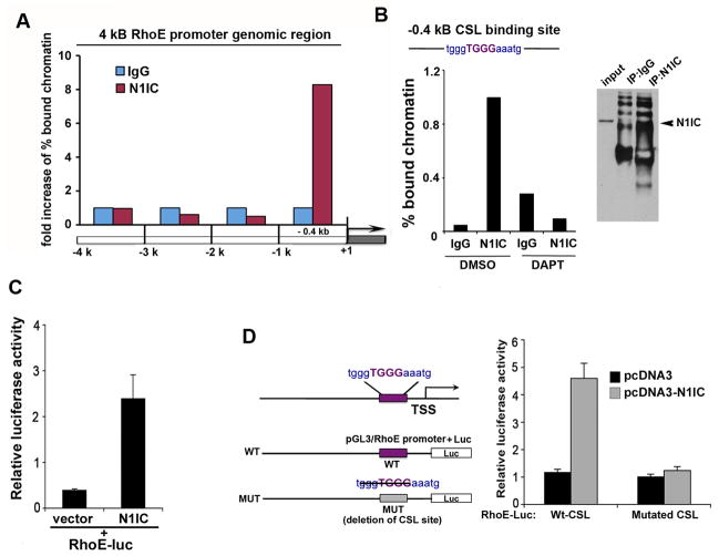
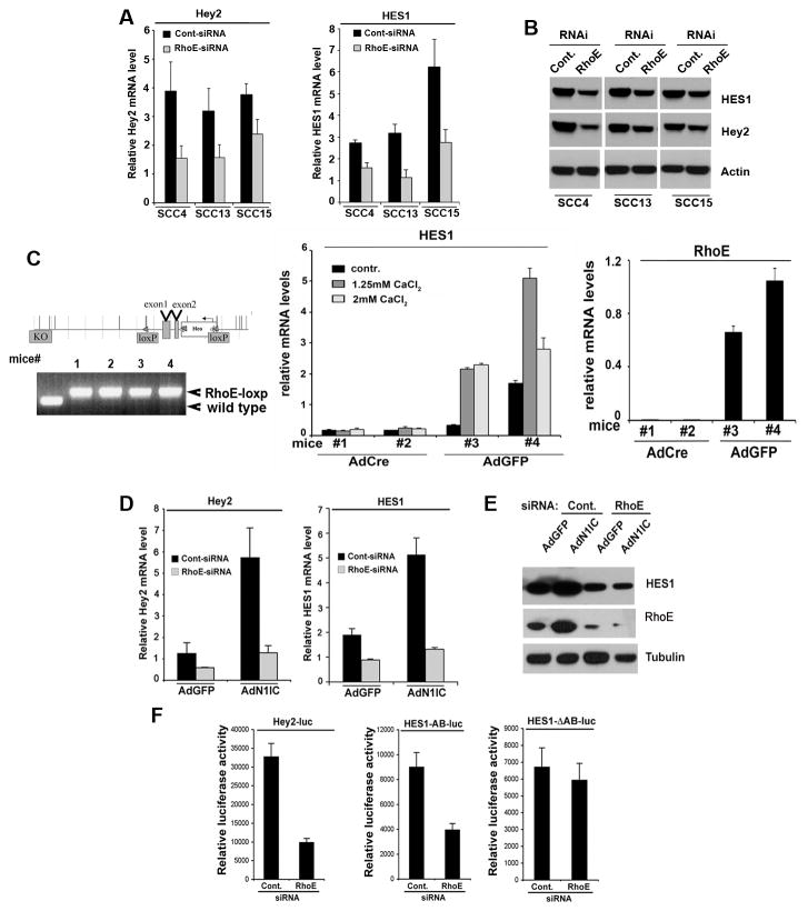
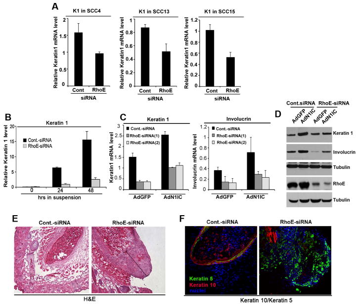
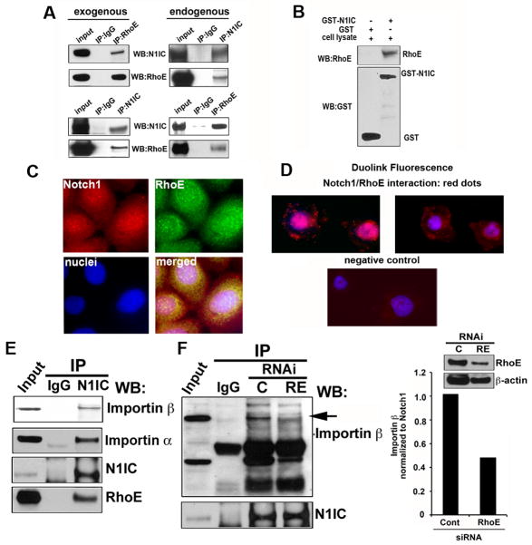
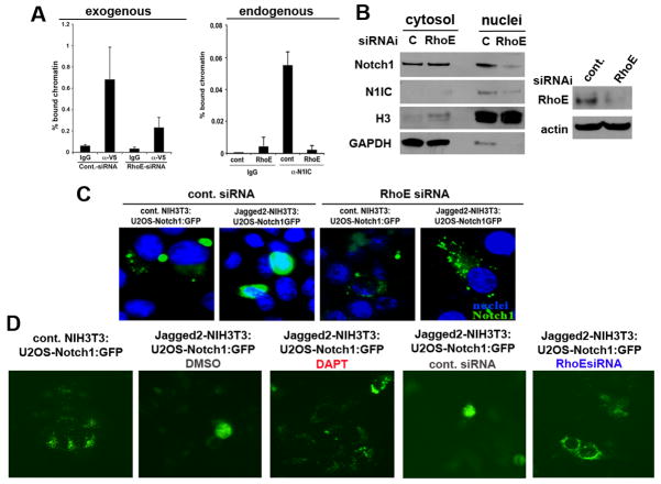
Aberrations of Notch signaling have been implicated in a variety of human cancers. Oncogenic mutations in NOTCH1 are common in human T-cell leukemia and lymphomas. However, loss-of-function somatic mutations in NOTCH1 arising in solid tumors imply a tumor suppressor function, which highlights the need to understand Notch signaling more completely. Here, we describe the small GTPase RhoE/Rnd3 as a downstream mediator of Notch signaling in squamous cell carcinomas (SCC) that arise in skin epithelia. RhoE is a transcriptional target of activated Notch1, which is attenuated broadly in SCC cells. RhoE depletion suppresses Notch1-mediated signaling in vitro, rendering primary keratinocytes resistant to Notch1-mediated differentiation and thereby favoring a proliferative cell fate. Mechanistic investigations indicated that RhoE controls a key step in Notch1 signaling by mediating nuclear translocation of the activated portion of Notch1 (N1IC) through interaction with importins. Our results define RhoE as a Notch1 target that is essential for recruitment of N1IC to the promoters of Notch1 target genes, establishing a regulatory feedback loop in Notch1 signaling. This molecular circuitry may inform distinct cell fate decisions to Notch1 in epithelial tissues, where carcinomas such as SCC arise.


Conflict of interest statement

The authors declare no competing interests.

Figures

Figure 1
Figure 1. RhoE expression is down-regulated in SCC cell lines and tumors in parallel with Notch1 levels
A, Primary HKC were analyzed in parallel with SCC cell lines (SCCO28, Cal27 and FaDu) by real time RT-PCR for Notch1 and RhoE mRNA levels using 36B4 as internal control. B, The same set of cells as in A, were analyzed for protein levels of Notch1 and RhoE using β-actin as a loading control. In addition protein levels of the cleaved part of Notch1 were detected with Val1744 specific antibody (lower panel). C, Total RNA from 23 surgically excised oral SCC subjected to laser dissection microscopy (as in (5) were analyzed along with normal epithelium for RhoE mRNA expression by real time RT-PCR using 36B4 as control. The results are expressed as fold changes of RhoE expression in the tumors versus normal epithelium. Error bars represent Standard Deviations, calculated based on triplicate values.
Figure 2
Figure 2. RhoE expression is regulated by Notch1 in keratinocytes
A, Notch activation induces RhoE expression. HKC were infected with AdN1IC or control AdGFP, followed by assessment of RhoE mRNA levels by real time RT-PCR (left panel) and protein levels after Western blot. B, HKC were co-cultured with control mouse NIH 3T3 fibroblasts or NIH 3T3 fibroblasts stably expressing full-length Jagged1 for 48 hours, followed by real-time RT-PCR analysis of RhoE mRNA levels. C, HKC were treated with 10 μM DAPT or DMSO vehicle control for 24 hrs, after that DAPT was washed out with PBS and cell culture medium and cells were treated with cycloheximide for 2. RhoE mRNA levels were then assessed by real time RT-PCR analysis. D, MAM51-mediated inhibition of Notch transcription suppresses RhoE expression. (E) Western blot analysis of RhoE protein levels in MAM51 overexpressing or DAPT treated primary HKC with tubulin as a loading control. F, Primary mouse keratinocytes from homozygous Notchloxp/loxp were infected with AdCre or AdGFP control. At 72 hours after infection, cells were analyzed either by real time RT-PCR analysis and immunoblotting for RhoE expression levels using 36B4 mRNA levels or γ-tubulin protein levels for normalization, respectively. The deletion of the Notch1 gene was confirmed by immunoblotting with a Notch specific antibody. G, Real time RT-PCR analysis was used to analyze RhoE mRNA levels, using mouse GAPDH for internal normalization. Relative mRNA levels are presented as a fold change as compared to the control condition. Error bars represent Standard Deviations, calculated based on three independent measurements.
Figure 3
Figure 3. Identification of RhoE as a transcriptional target of Notch1
A, Endogenous Notch1 is enriched at −1kb genomic region from the transcription start site (TSS) of human RhoE. Primary HKC were forced to differentiate in suspension and processed for scanning ChIP of the −5kb genomic region upstream of the human RhoE TSS, as described in the Supplementary Information, using an antibody against the activated form of Notch1 or purified rabbit IgGs. B, Differentiated primary HKC, teraetd with DMSO control or DAPT were processed for ChIP as in (A) followed by real time PCR analysis (upper panel) or conventional PCR (lower pane) using specific primers (described in Supplementary Information) to amplify the −0.4kb region of the human RhoE promoter containing the CSL binding site. Efficiency of the immunoprecipitation is shown in the right panel: Western blot analysis of immunoprecipitates performed either with non-specific IgG or with the antibody against the activated form of Notch1. C, SCC028 cells were transfected with a N1IC construct or empty vector together with the RhoE reporter (RhoE-luc) and phRL-TK Renilla reporter. D, The promoter regions were analyzed using the rVista software for identification of transcription factor binding sites. The CSL binding site at position −0.4 kb was deleted, and SCC028 keratinocyte cells were transfected with the mutated or with the wild type luciferase reporter constructs together with the NICD construct and phRL-TK Renilla reporter. Error bars represent Standard Deviations, calculated based on three independent measurements.
Figure 4
Figure 4. Depletion of RhoE inhibits downstream Notch signaling
A, SCC4, SCC13 and SCC15 cancer cell lines were transfected with control and RhoE siRNA and the levels of the Notch target genes Hey2 and HES1 were analyzed by real time RT-PCR analysis using 36B4 as an internal control. B, Protein levels of Hey2, HES1 and RhoE in the same cells were analyzed by Western blot using actin levels as an internal loading control. C, Inducible keratinocyte-specific deletion of the RhoE gene and resulting effects on Notch signaling and keratinocyte differentiation. Left upper panel: Schematic representation of the murine RhoE gene and of the expected recombination products leading to its inducible tissue-specific deletion. After induction of Cre recombinase activity (the loxP sites are indicated with red arrows), the genomic portion harboring the first two exons are deleted. Left lower panel: PCR analysis of genomic DNA of offspring from the RhoE loxp/loxp homozygous mice: the primers amplify a larger fragment including the loxp site and wild type genomic DNA is presented as a control. Middle panel: Real-time RT-PCR analysis of mRNA isolated from primary keratinocytes derived from mutant RhoE loxp/loxp mice and infected with an adenovirus expressing the Cre recombinase or an Ad-GFP control. At 2 days after infection levels of Hes1 are decreased in cells with Cre activity upon calcium mediated induction of differentiation. Depletion of RhoE mRNA levels shown on the right. D, Primary HKC under growing adherent conditions were transfected with control (scrambled siRNA) or RhoE siRNA, and 48 hours later were infected with adenoviruses expressing either GFP (AdGFP) or the activated form of Notch1 (AdN1IC). 24 hours later expression of Notch1 target genes, Hey2 and HES1 was analyzed by real time RT-PCR. E, RhoE and HES1 expression in the same cells was analyzed by Western blot. F, SCC028 cells transfected with control or RhoE siRNA were transfected with luciferase reporters for Hey2 (Hey2-luc, 0.5mg), wild type HES1 containing both CSL binding sites in the promoter region (HES1-AB-luc, 0.5mg) or HES1-ΔAB-luc with mutated CSL binding sites together with a Renilla minimal reporter (0.05 mg) for internal normalization. 24 hrs after transfection, luciferase activity was measured and normalized with Renilla activity. Error bars represent Standard Deviations, calculated based on three independent measurements.
Figure 5
Figure 5. RhoE ablation suppresses growth and commitment to differentiation in vitro and enhances the proliferative phenotype in vivo
A, SCC4, SCC13 and SCC15 cells were depleted of RhoE expression using siRNA and Keratin 1 mRNA was analyzed 48 hrs later using real time RT-PCR. B, Primary HKC transfected with control or RhoE siRNA were kept under growing conditions (time point 0) or induced to differentiate by suspension or the indicated times. Keratin1 mRNA levels were analyzed by real time RT-PCR. C, Knockdown of RhoE inhibits the Notch1-mediated induction of Keratin 1 and Involucrin. Primary HKC under growing adherent conditions were transfected with control or two different RhoE siRNAs, and 48 hours later infected with adenoviruses AdGFP or AdN1IC. 24 hours later commitment of keratinocytes to differentiation was analyzed by Keratin 1 or Involucrin mRNA levels. D, HKC transfected with control or RhoE siRNA were treated as in C, and analyzed for RhoE, Keratin 1 and Involucrin protein levels by Western blot using tubulin as a loading control. E, Human HKC transfected with control or RhoE siRNA were injected into immunodeficient nude mice at the dermal-epidermal junction. To minimize individual animal variations, the same mice were injected in parallel with control siRNA transfected keratinocytes. 7 days later cysts were harvested, and tissues were processed. F, The same cysts from above experiments were stained with a Keratin 5 specific antibody (green staining in the right panel). Shown are multilayer cysts with well-differentiated keratinocytes building a cornified envelope in the control samples and a large “basal” layer of proliferating keratinocytes with increased Keratin 5 expression (green staining) and decreased Keratin 10 expression (red staining) in the RhoE siRNA tissues. Nuclei were stained in blue (Hoechst staining). Error bars represent Standard Deviations, calculated based on three independent measurements.
Figure 6
Figure 6. RhoE binds to N1IC and promotes its importin β1-mediated nuclear import
A, Coimmunoprecipitation of N1IC by RhoE. Cellular extracts from U2OS cells stably expressing Flag-tagged RhoE and infected with AdN1IC were immunoprecipitated using Flag antibody followed by Western blotting for N1CD and RhoE. Endogenous association between N1IC and RhoE in SCC4 cells was analyzed be coimunoprecipitation of N1IC or RhoE followed by Western blot for the indicated proteins. B, GST-pull down assay was also performed: RhoE binds to recombinant N1IC fused to GST but not to GST alone. C, N1IC and RhoE colocalize in the cytoplasm and the nucleus of SCC4 cells by immunofluorescence staining using antibodies for endogenous RhoE and N1IC. The images were then analyzed by confocal microscopy. D, Representative images of SCC4 cell with Duolink fluorescence (red spots) generated by the interaction of the anti RhoE and anti N1IC antibodies (DAPI was used to stain the nucleus). Negative control images were generated by omitting the RhoE antibody. E, N1IC interacts with Importin family members. Cellular lysates of SCC4 cells were tested for the endogenous association between N1IC and importin β1 or α by immunoprecipitation with anti N1IC versus control IgG followed by Western blotting for importin α and β1. F. Depletion of RhoE abrogates the binding between NIIC and importin β1. SCC4 cells were transfected with either scrambled or RhoE siRNA and 48 hours later subjected to immunoprecipitation with an anti N1IC antibody followed by Western blotting using anti importin β1 and Notch1 antibodies. Rabbit IgG was used as a control (left panel). The decrease of immunoprecipitaed importin β1 was measured using UNISCANIT software. RhoE knock-down was measured by immunoblotting (right panel).
Figure 7
Figure 7. RhoE promotes N1IC transcriptional activity and importin β1-mediated nuclear import
A, RhoE knock-down inhibits Notch1-induced CSL-transcription in normal keratinocytes. Human primary keratinocytes under growing conditions were transfected with control siRNA, or RhoE siRNA followed by infection with an adenoviral construct encoding the activated form of Notch1 with a V5 C-terminal tag. 24 hours later cells were processed for ChIP with V5 specific antibodies or non-immune IgG control followed by PCR amplification of the CSL/Notch binding site-containing region of the human HES1 promoter (left panel). Differentiated primary HKC were processed for ChIP as in Figure 3 using endogenous N1IC for immunoprecipitation (right panel). The results from the ChIP assay were quantified by real time PCR analysis. Error bars represent Standard Deviations, calculated based on three independent measurements. B, RhoE inhibition prevents accumulation of activated Notch1 in the nucleus. Primary HKC transfected with RhoE siRNA were induced to differentiate by suspension culture, followed by nuclear and cytoplasmic fractionation and immunoblot analysis for the full length and activated Notch1 proteins using H3 and GAPDH as loading controls for the nuclear and cytosolic fractions, respectively. RhoE knockdown was assessed by Western blot analysis as shown in the right panel. C, U2OS cells stably expressing the full length Notch1-GFP (the GFP tag is at the intracellular end of the protein) transfected with control or RhoE siRNA and seeded over NIH3T3 cells expressing control empty vector or full length Jagged2 were analyzed for ligand mediated translocation of cleaved Notch1-GFP to the nucleus 24hrs later by fluorescent microscopy. D, Same co-culture system as in C but imaging was performed on live cells and the effects of RhoE siRNA were compared to DAPT treatment.

References

    1. Devgan V, Mammucari C, Millar SE, Brisken C, Dotto GP. p21WAF1/Cip1 is a negative transcriptional regulator of Wnt4 expression downstream of Notch1 activation. Genes & development. 2005;19:1485–95. - PMC - PubMed
    1. Nguyen BC, Lefort K, Mandinova A, Antonini D, Devgan V, Della Gatta G, et al. Cross-regulation between Notch and p63 in keratinocyte commitment to differentiation. Genes & development. 2006;20:1028–42. - PMC - PubMed
    1. Rangarajan A, Talora C, Okuyama R, Nicolas M, Mammucari C, Oh H, et al. Notch signaling is a direct determinant of keratinocyte growth arrest and entry into differentiation. The EMBO journal. 2001;20:3427–36. - PMC - PubMed
    1. Kolev V, Mandinova A, Guinea-Viniegra J, Hu B, Lefort K, Lambertini C, et al. EGFR signalling as a negative regulator of Notch1 gene transcription and function in proliferating keratinocytes and cancer. Nat Cell Biol. 2008;10:902–11. - PMC - PubMed
    1. Lefort K, Mandinova A, Ostano P, Kolev V, Calpini V, Kolfschoten I, et al. Notch1 is a p53 target gene involved in human keratinocyte tumor suppression through negative regulation of ROCK1/2 and MRCKalpha kinases. Genes & development. 2007;21:562–77. - PMC - PubMed

Publication types