. 2014;127(6):861-79.
doi: 10.1007/s00401-014-1256-4. Epub 2014 Feb 14.

Next-generation active immunization approach for synucleinopathies: implications for Parkinson's disease clinical trials

Affiliations

Next-generation active immunization approach for synucleinopathies: implications for Parkinson's disease clinical trials

Markus Mandler et al. Acta Neuropathol. 2014.

Abstract

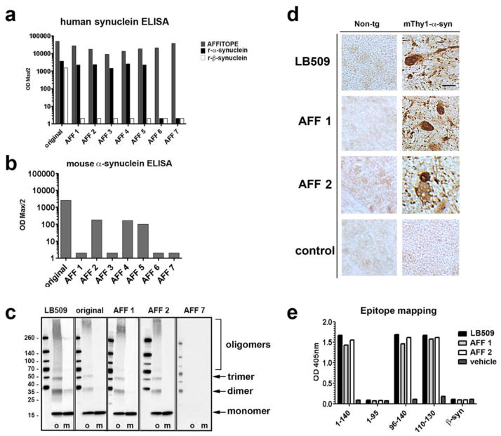
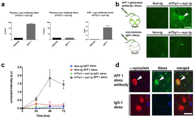
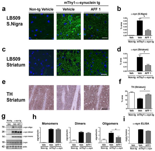
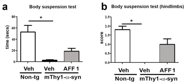
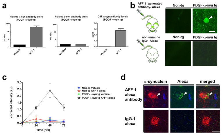
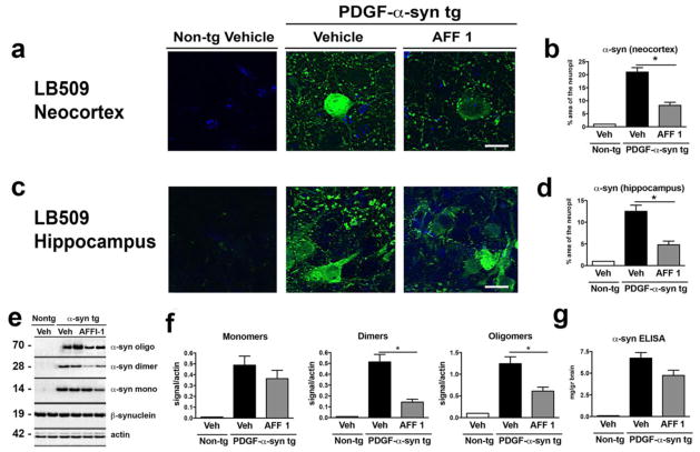
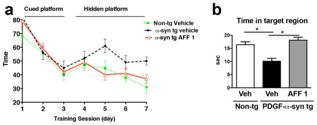
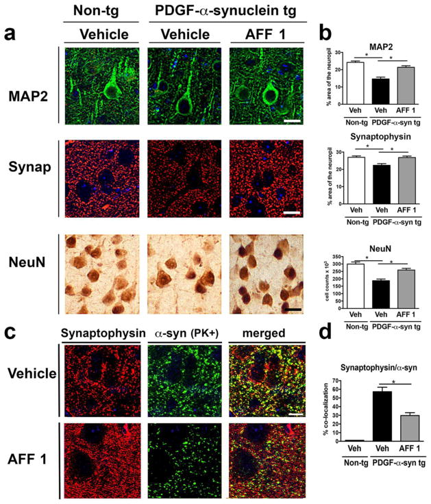
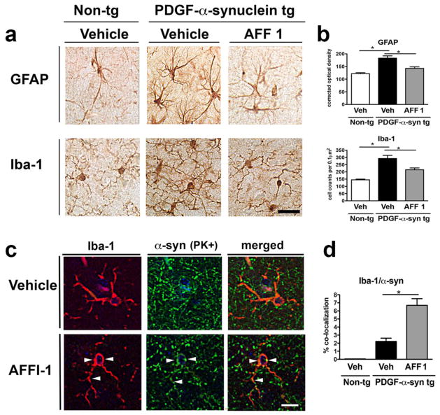
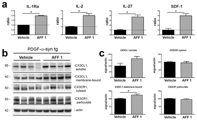
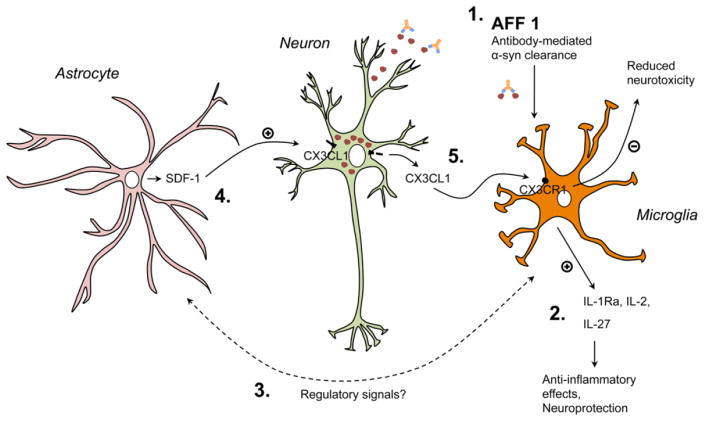
Immunotherapeutic approaches are currently in the spotlight for their potential as disease-modifying treatments for neurodegenerative disorders. The discovery that α-synuclein (α-syn) can transmit from cell to cell in a prion-like fashion suggests that immunization might be a viable option for the treatment of synucleinopathies. This possibility has been bolstered by the development of next-generation active vaccination technology with short peptides-AFFITOPEs(®) (AFF)- that do not elicit an α-syn-specific T cell response. This approach allows for the production of long term, sustained, more specific, non-cross reacting antibodies suitable for the treatment of synucleinopathies, such as Parkinson's disease (PD). In this context, we screened a large library of peptides that mimic the C-terminus region of α-syn and discovered a novel set of AFF that identified α-syn oligomers. Next, the peptide that elicited the most specific response against α-syn (AFF 1) was selected for immunizing two different transgenic (tg) mouse models of PD and Dementia with Lewy bodies, the PDGF- and the mThy1-α-syn tg mice. Vaccination with AFF 1 resulted in high antibody titers in CSF and plasma, which crossed into the CNS and recognized α-syn aggregates. Active vaccination with AFF 1 resulted in decreased accumulation of α-syn oligomers in axons and synapses, accompanied by reduced degeneration of TH fibers in the caudo-putamen nucleus and by improvements in motor and memory deficits in both in vivo models. Clearance of α-syn involved activation of microglia and increased anti-inflammatory cytokine expression, further supporting the efficacy of this novel active vaccination approach for synucleinopathies.


Conflict of interest statement

CONFLICT OF INTEREST

The authors Markus Mandler, Harald Weninger, Radmila Santic, Stefanie Meindl, Benjamin Vigl, Oskar Smrzka and Achim Schneeberger are employees of AFFiRiS, the company that commercialize the AFFITOPEs® described in the manuscript. The author Frank Mattner is co-founder of AFFiRiS. The authors Elvira Valera, Edward Rockenstein, Christina Patrick, Anthony Adame and Eliezer Masliah declare that they have no conflict of interest.

Figures

Fig. 1
Fig. 1. Immune response of mice vaccinated with AFFITOPEs®
BALB/c mice were vaccinated three times and the titer of antibodies against different peptides was assessed by peptide ELISA. (a) Titer of antibodies against the injected peptide (AFFITOPE®) as well as against recombinant full-length human α-syn or β-syn. The original C-terminus of human α-syn (original, 110–130) was used as control. Antibody titers are depicted as ODmax/2. (b) Titer of antibodies against recombinant full-length murine α-syn. The original C-terminus of human α-syn (original, 110–130) was used as control. Antibody titers are depicted as ODmax/2. (c) Detection of oligomeric (o) and monomeric (m) α-syn by immunoblotting using plasma of single-vaccinated non-tg mice. The α-syn-specific antibody LB509 was used as positive control. (d) AFF 1 or AFF 2-induced antibodies were used to recognize human α-syn in brain sections of non-tg mice and of mThy1-α-syn tg mice. LB509 was used as positive control. Scale bar = 5 μm. (e) Epitope mapping of AFF 1 and AFF 2-induced antibodies. LB509 was used as control antibody. Maximum OD at a dilution of 1:100 is depicted
Fig. 2
Fig. 2. Reactivity of antibodies generated after vaccination with AFF 1 in mThy1-α-syn tg mice
(a) Plasma or CSF of mThy1-α-syn mice treated with AFF 1 or vehicle were analyzed for the presence of α-syn or β-syn-specific antibodies after repeated vaccinations. Antibody titers are depicted as ODmax/2. (b) AFF 1-induced antibodies were tagged with Alexa-488 and administered to non-tg or mThy1-α-syn mice. AFF 1-induced antibodies bound α-syn in neuronal bodies (arrow-head) and neuropil. As negative control, a non-immune IgG1 was used and no binding was observed. Scale bar = 5 μm. (c) AFF 1-induced antibodies or non-immune IgG were tagged with Alexa-488 and administered to non-tg or mThy1-α-syn mice. A time course analysis was performed, showing that green fluorescence was only increased in brain sections of mThy1-α-syn tg animals injected with Alexa-488-tagged AFF 1 induced antibodies. Results are shown as corrected intensity values. (d) AFF 1-induced antibodies were tagged with Alexa-488 and used for immunofluorescence staining of brain sections of mThy1-α-syn tg mice (green), together with an antibody against α-syn (red). Colocalization was observed in neuronal cell bodies (arrow-head). As negative control, non-immune IgG1 was used and no α-syn staining was observed. Scale bar = 5 μm. Results are expressed as average ± SEM
Fig. 3
Fig. 3. Immunization with AFF 1 reduced α-syn load in mThy1-α-syn tg mice
α-syn levels were measured in non-tg mice and mThy1-α-syn tg mice immunized either with vehicle or AFF 1. (a) α-syn immunostaining of substantia nigra using the α-syn antibody LB509 (green). Cell nuclei were stained with DAPI (blue). Scale bar = 10 μm. (b) Quantification of the percentage of neuropil area positive for α-syn in substantia nigra. (c) α-syn immunostaining of striatum using the α-syn antibody LB509 (green). Cell nuclei were stained with DAPI (blue). Scale bar = 10 μm. (d) Quantification of the percentage of neuropil area positive for α-syn in striatum. (e) Tyrosine hydroxylase (TH) immunostaining of striatum. Scale bar = 10 μm. (f) Quantification of the percentage of neuropil area positive for TH in striatum. (g) Immunoblot analysis of α-syn species (oligomers, dimers, and monomers). Levels of β-syn did not change with any of the treatments. β-actin was used as loading control. (h) Densitometric analysis of immunoblot results. (i) ELISA analysis of total levels of human α-syn. Results are expressed as average ± SEM. (*) p<0.05
Fig. 4
Fig. 4. Effect of vaccination with AFF 1 on motor dysfunction in mThy1-α-syn tg mice
Non-tg mice and mThy1-α-syn tg mice immunized either with vehicle or AFF 1 were analyzed for motor dysfunction using the body suspension test at 9 months of age. (a) Latency to fall measured as the time the mouse is suspended until it falls from the bar. (b) The ability to use the hindlimbs was quantified using a three-category scale: a score of 0 indicates the inability to use the hindlimbs, 1 reflects the ability to use one hindlimb, and a score of 2 indicates the use of both hindlimbs to support the body. Results are expressed as average ± SEM. (*) p<0.05
Fig. 5
Fig. 5. Reactivity of antibodies generated after vaccination with AFF 1 in PDGF-α-syn tg mice
(a) Plasma or CSF of PDGF-α-syn mice treated with AFF 1 or vehicle were analyzed for the presence of α-syn or β-syn-specific antibodies after repeated vaccinations. Antibody titers are depicted as ODmax/2. (b) AFF 1-induced antibodies were tagged with Alexa-488 and administered to non-tg or PDGF-α-syn mice. AFF 1-induced antibodies bounded α-syn in neuronal bodies (arrow-head) and neuropil. As negative control, a non-immune IgG1 was used and no binding was observed. Scale bar = 5 μm. (c) AFF 1-induced antibodies or non-immune IgG were tagged with Alexa-488 and administered to non-tg or PDGF-α-syn mice. A time course analysis was performed, showing that green fluorescence was only increased in brain sections of PDGF-α-syn tg animals injected with Alexa-488-tagged AFF 1 induced antibodies. Results are shown as corrected intensity values. (d) AFF 1-induced antibodies were tagged with Alexa-488 and used for immunofluorescence staining of brain sections of PDGF-α-syn tg mice (green), together with an antibody against α-syn (red). Colocalization was observed in neuronal cell bodies (arrow-head). As negative control, non-immune IgG1 was used and no α-syn staining was observed. Scale bar = 5 μm. Results are expressed as average ± SEM
Fig. 6
Fig. 6. Immunization with AFF 1 reduced α-syn load in PDGF-α-syn tg mice
α-syn levels were measured in non-tg mice and PDGF-α-syn tg mice immunized either with vehicle or AFF 1. (a) α-syn immunostaining of neocortex using the α-syn antibody LB509 (green). Cell nuclei were stained with DAPI (blue). Scale bar = 5 μm. (b) Quantification of the percentage of neuropil area positive for α-syn in neocortex. (c) α-syn immunostaining of hippocampus using the α-syn antibody LB509 (green). Cell nuclei were stained with DAPI (blue). Scale bar = 5 μm. (d) Quantification of the percentage of neuropil area positive for α-syn in hippocampus. (e) Immunoblot analysis of α-syn species (oligomers, dimers, and monomers). Levels of β-syn did not change with any of the treatments. β-actin was used as loading control. (f) Densitometric analysis of immunoblot results. (I) ELISA analysis of total levels of human α-syn. Results are expressed as average ± SEM. (*) p<0.05
Fig. 7
Fig. 7. Effect of vaccination with AFF 1 on memory deficits in PDGF-α-syn tg mice
Non-tg mice or PDGF-α-syn tg mice were immunized either with vehicle or AFF 1, and spatial memory was measured using the water maze. (a) Time elapsed for finding the platform (escape latency), which was either cued (day 1–3) or hidden (day 4–7). (b) Time spent by the mice in the platform quadrant (target region) during the probe test. Results are expressed as average ± SEM. (*) p<0.05
Fig. 8
Fig. 8. Effect of vaccination with AFF 1 on the synaptic pathology of PDGF-α-syn tg mice
(a) Immunostaining of synaptic (MAP2, green; synaptophysin, red) and neuronal markers (NeuN) in brain sections of non-tg mice or PDGF-α-syn tg mice immunized either with vehicle or AFF 1. Scale bar = 5 μm. (b) Quantification of the percentage of the MAP2- or synaptophysin-positive area of neuropil, and NeuN cell counts. (c) Double immunostaining for synaptophysin (red) and proteinase K (PK)-resistant α-syn (green) in vehicle and AFF 1-treated PDGF-α-syn tg mice. Scale bar = 5 μm. (d) Quantification of the percentage of co-localization between synaptophysin and PK-resistant α-syn. Results are expressed as average ± SEM. (*) p<0.05
Fig. 9
Fig. 9. Effect of vaccination with AFF 1 on astrogliosis and microgliosis in PDGF-α-syn tg mice
Non-tg mice or PDGF-α-syn tg mice were immunized either with vehicle or AFF 1, and glial markers were analyzed by immunohistochemistry. (a) Immunostaining of the astroglial marker GFAP and the microglial marker Iba1. Scale bar = 25 μm. (b) Quantification of the intensity of GFAP staining and Iba1 cell counts. (c) Double immunostaining for Iba1 (red) and proteinase K (PK)-resistant α-syn (green) in vehicle and AFF 1 treated PDGF-α-syn tg mice. Co-localization between both markers is denoted by arrow-heads. Scale bar = 5 μm. (d) Quantification of the percentage of colocalization between Iba1 and PK-resistant α-syn. Results are expressed as average ± SEM. (*) p<0.05
Fig. 10
Fig. 10. Effect of vaccination with AFF 1 on cytokine levels in PDGF-α-syn tg mice
(a) PDGF-α-syn tg mice were immunized either with vehicle or AFF 1, and cytokine levels were analyzed using a mouse cytokine array. Significant changes were observed for the cytokines IL-1Ra, IL-2, IL-27 and SDF-1. Results are expressed as densitometry analysis relative to the control condition of vehicle-treated PDGF-α-syn tg mice. (b) Immunoblot analysis of fractalkine (CX3CL1) and fractalkine receptor (CX3CR1) levels in the soluble and insoluble fractions of brain homogenates of PDGF-α-syn tg mice immunized either with vehicle or AFF 1. β-actin was used as loading control. (c) Densitometric analysis of the immunoblot results. Results are expressed as average ± SEM. (*) p<0.05
Fig. 11
Fig. 11. Possible anti-inflammatory mechanisms elicited by immunization with AFF 1 in α-syn tg mice
Microglial cells of the α-syn tg mice immunized with AFF 1 would clear out antibody-α-syn complexes, reducing its extracellular toxicity (1). Additionally, reduced levels of α-syn and regulatory signals would stimulate glial cells to increase the production of anti-inflammatory cytokines, such as IL-1Ra, IL-2, IL-27 (2) and SDF-1 (3). SDF-1 regulates fractalkine (CX3CL1) levels in neurons and induces its cleavage from the neuronal membrane (4). Soluble fractalkine would then interact with its receptor (CX3CR1) in microglial cells, inducing a reduction in microglial-dependent neurotoxicity (5). However, the precise molecular mechanisms by which immunization with AFF 1 would induce anti-inflammatory cytokine production remain to be studied. Red spheres, α-syn oligomers

References

    1. Amor S, Puentes F, Baker D, van der Valk P. Inflammation in neurodegenerative diseases. Immunology. 2010;129 (2):154–169. doi: 10.1111/j.1365-2567.2009.03225.x. - DOI - PMC - PubMed
    1. Amschl D, Neddens J, Havas D, Flunkert S, Rabl R, Romer H, Rockenstein E, Masliah E, Windisch M, Hutter-Paier B. Time course and progression of wild type alpha-synuclein accumulation in a transgenic mouse model. BMC Neurosci. 2013;14:6. doi: 10.1186/1471-2202-14-6. - DOI - PMC - PubMed
    1. Angot E, Brundin P. Dissecting the potential molecular mechanisms underlying alpha-synuclein cell-to-cell transfer in Parkinson’s disease. Parkinsonism Relat Disord. 2009;15(Suppl 3):S143–147. doi: 10.1016/S1353-8020(09)70802-8. - DOI - PubMed
    1. Aquilano K, Baldelli S, Rotilio G, Ciriolo MR. Role of nitric oxide synthases in Parkinson’s disease: a review on the antioxidant and anti-inflammatory activity of polyphenols. Neurochem Res. 2008;33 (12):2416–2426. doi: 10.1007/s11064-008-9697-6. - DOI - PubMed
    1. Asuni AA, Boutajangout A, Quartermain D, Sigurdsson EM. Immunotherapy targeting pathological tau conformers in a tangle mouse model reduces brain pathology with associated functional improvements. J Neurosci. 2007;27 (34):9115–9129. doi: 10.1523/JNEUROSCI.2361-07.2007. - DOI - PMC - PubMed

Publication types

MeSH terms