Review
. 2014 Feb 14;114(4):717-29.
doi: 10.1161/CIRCRESAHA.114.301863.

Matrix revisited: mechanisms linking energy substrate metabolism to the function of the heart

Affiliations
Review

Matrix revisited: mechanisms linking energy substrate metabolism to the function of the heart

Andrew N Carley et al. Circ Res. .

Abstract

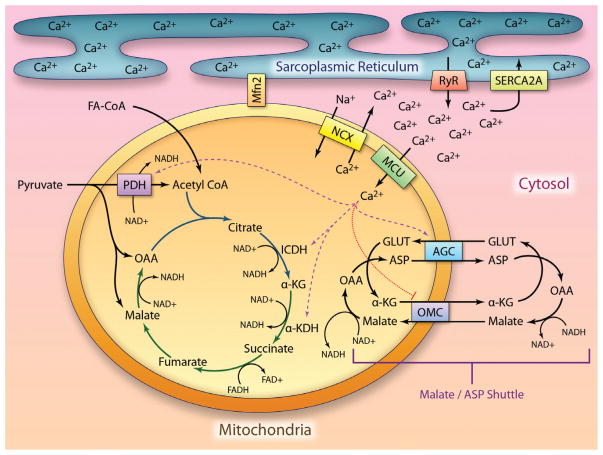
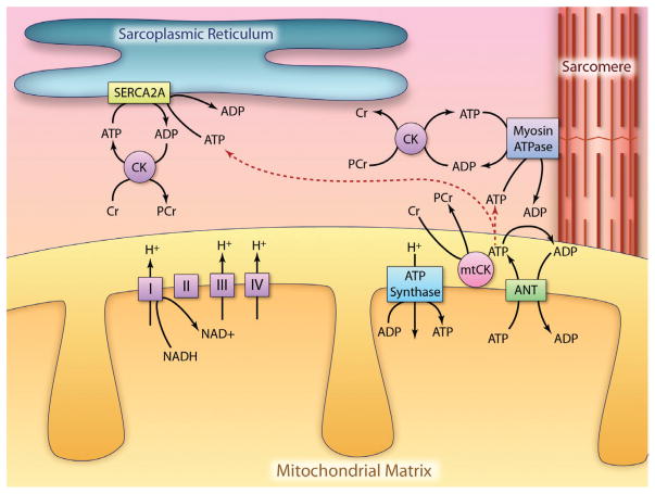
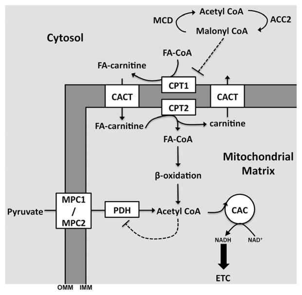
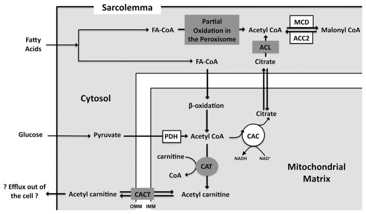
Metabolic signaling mechanisms are increasingly recognized to mediate the cellular response to alterations in workload demand, as a consequence of physiological and pathophysiological challenges. Thus, an understanding of the metabolic mechanisms coordinating activity in the cytosol with the energy-providing pathways in the mitochondrial matrix becomes critical for deepening our insights into the pathogenic changes that occur in the stressed cardiomyocyte. Processes that exchange both metabolic intermediates and cations between the cytosol and mitochondria enable transduction of dynamic changes in contractile state to the mitochondrial compartment of the cell. Disruption of such metabolic transduction pathways has severe consequences for the energetic support of contractile function in the heart and is implicated in the pathogenesis of heart failure. Deficiencies in metabolic reserve and impaired metabolic transduction in the cardiomyocyte can result from inherent deficiencies in metabolic phenotype or maladaptive changes in metabolic enzyme expression and regulation in the response to pathogenic stress. This review examines both current and emerging concepts of the functional linkage between the cytosol and the mitochondrial matrix with a specific focus on metabolic reserve and energetic efficiency. These principles of exchange and transport mechanisms across the mitochondrial membrane are reviewed for the failing heart from the perspectives of chronic pressure overload and diabetes mellitus.

Keywords: cytosol; diabetes mellitus; heart failure; metabolic pathways; metabolism; mitochondria; mobilization.


Figures

Figure 1
Figure 1. Calcium regulation of mitochondrial metabolism
Calcium (Ca2+) released by the sarcoplasmic reticulum is taken up by the mitochondria via the mitochondrial calcium uniporter (MCU). Mitochondria are found in close association with the calcium release channels in the sarcoplasmic reticulum, the ryanodine receptors (RyR), creating a Ca2+ microdomain. Mitofusin2 (Mfn2) acts to bring the mitochondria and sarcoplasmic reticulum (SR) into close communication. Ca2+ activates (designated by the dashed purple lines) mitochondrial metabolism by increasing the activities of pyruvate dehydrogenase (PDH), isocitrate dehydrogenase (ICDH), and α-ketoglutarate (α-KG) dehydrogenase (α-KDH) resulting in increased metabolism of pyruvate and fatty acyl CoA and increased NADH formation. Ca2+ also increases the exchange rate of malate/aspartate (Malate/ASP) shuttle by increasing the activity of the ASP/glutamate (GLUT) exchangers 1 and 2 (AGC1/2), while inhibiting (indicated by the dotted red line) net efflux of citric acid cycle (CAC) intermediates out of the mitochondria via the oxaloacetate (OAA)/malate cotransporter (OMC). The malate/ASP shuttle is responsible for maintaining the cytoplasmic NAD+ pool. The 2 spans of the CAC are indicated by blue (reductive span) and green (oxidative span) lines. Pyruvate can feed into the reductive span through PDH or feed into OAA and malate pools in the oxidative span via anaplerotic flux. Ca2+ is cleared from the mitochondrial matrix by the actions of the Na+/Ca2+ exchanger (NCX). The magnitude of the calcium release by the SR is dependent on the actions of SR Ca2+-ATPase (SERCA2a), which facilitates Ca2+ reuptake into the SR (illustration credit: Ben Smith). FA-CoA indicates fatty acyl-carnitine.
Figure 2
Figure 2. Phosphotransfer systems in the heart
ATP is formed within the mitochondria by ATP synthase, which uses the proton gradient across the inner mitochondrial membrane (IMM) to catalyze ATP formation from ADP and Pi (not depicted). The proton gradient is generated by actions of the electron transport chain (shown as complexes I–IV) after donation of an electron by NADH at complex I. Once formed ATP can cross the IMM via the adenine nucleotide transporter (ANT). ANT also brings ADP into the mitochondria from the cytoplasm. The energy contained within ATP is then transferred to the cytoplasmic phosphocreatine (PCr) pool through the actions of a mitochondrial isoform of creatine kinase (CK). PCr is less diffusion limited than ATP and is therefore an efficient means of transmitting energy to the peripheral sites of ATP utilization, depicted as sarcoplasmic reticulum (SR) Ca2+-ATPase (SERCA2a)–dependent calcium reuptake by the sarcoplasmic reticulum and myosin ATPase-dependent cross-bridge cycling within the sarcomere. PCr is converted back to free creatine (Cr) by cytoplasmic CK with the freed energy used for ATP formation and driving the peripheral ATPases. An alternative phosphotransfer pathway (depicted by a dashed line) is for ATP to directly travel from the mitochondria to the sites of utilization independent of the PCr:CK system. As ATP is diffusion limited, the direct transfer of ATP from the mitochondria to a site of utilization requires close localization between the mitochondria and the peripheral ATPase (illustration credit: Ben Smith).
Figure 3
Figure 3. Protein transporters regulate metabolic substrate entry into the mitochondrial matrix and control the relative rates of oxidative metabolism
Fatty acyl CoA (FA-CoA) and pyruvate enter the mitochondria through protein transporters. FA-CoA is first converted to fatty acyl-carnitine (FA-carnitine) by carnitine palmitoyltransferase 1 (CPT1) located on the outer mitochondrial membrane (OMM). CPT1 activity is controlled by the concentration of malonyl CoA and therefore the activities of malonyl CoA decarboxylase (MCD) and acetyl CoA carboxylase 2 (ACC2) indirectly regulate CPT1 activity. FA-carnitine enters the mitochondria via carnitine-acylcarnitine translocase where it is then converted back to FA-CoA by CPT2 located on the inner mitochondrial membrane (IMM). FA-CoA is then able to undergo oxidation within the mitochondria to yield acetyl CoA for the citric acid cycle (CAC). Pyruvate enters the mitochondria via the newly discovered mitochondrial pyruvate carriers 1 and 2 (MPC1 and MPC2) protein complex. Pyruvate is then converted to acetyl CoA by the actions of pyruvate dehydrogenase (PDH). Acetyl CoA exerts product inhibition on the activity of PDH. CACT indicates carnitine-acylcarnitine translocase.
Figure 4
Figure 4. Intermediate exchange across the mitochondrial membrane buffers the cytoplasmic and mitochondrial acetyl CoA concentrations
The concentration of acetyl CoA formed during both pyruvate and fatty acyl CoA (FA-CoA) oxidation is buffered within the mitochondrial matrix by carnitine acetyltransferase (CAT). CAT converts acetyl CoA to acetyl carnitine. Acetyl carnitine can then be exported out of the mitochondria by carnitine-acylcarnitine translocase and potentially be cleared from the cell into the circulation or re-enter the mitochondria to undergo oxidative metabolism. Cytoplasmic acetyl CoA does not originate from mitochondrial acetyl CoA but rather from citric acid cycle (CAC) intermediate citrate, which is converted to acetyl CoA via ATP citrate lyase (ACL). Partial oxidation of FA-CoA in the peroxisome has also been suggested to contribute to the cytoplasmic acetyl CoA pool. The activities of malonyl CoA decarboxylase (MCD) and acetyl CoA carboxylase 2 (ACC2) regulate the ratio of cytoplasmic acetyl CoA to malonyl CoA. CACT indicates carnitine-acylcarnitine translocase; IMI, inner mitochondrial membrane; and OMM, outer mitochondrial membrane.
Figure 5
Figure 5. Turnover of long-chain fatty acids (LCFAs) within the myocardial triacylglyceride (TAG) pool and their importance in myocardial signaling
LCFAs, found within the circulation either complexed to albumin or esterified into triacylglyceride rich lipoproteins (TG-lipoprotein), enter the cell via a protein-mediated mechanism across the sarcolemma that is sensitive to CD36 expression. The insulin sensitive fatty acid transport protein 1 (FATP1) and the cardiac-specific FATP6 have also been implicated in LCFA uptake by the heart; however, at present only a direct effect of CD36 expression on TAG dynamics has been demonstrated. Lipoprotein lipase (LPL) is required to liberate LCFA from their esterified form in TG-lipoprotein. After entry into the myocyte LCFA can either enter the mitochondria for oxidation or cycle through the TAG pool (TAG turnover). TAG turnover is defined by the diacylglycerol acyltransferase 1 (DGAT1) dependent on rate (esterification) and the adipose triglyceride lipase (ATGL) dependent on off rate (lipolysis). After LCFA cycling through the TAG pool, LCFA can then be transported into the mitochondria for oxidation or used to initiate gene transcription via peroxisome proliferator–activated receptor α (PPARα). LCFA must first be cycled through the TAG pool to efficiently activate gene transcription.

Similar articles

Cited by

References

    1. Balaban RS. Cardiac energy metabolism homeostasis: role of cytosolic calcium. J Mol Cell Cardiol. 2002;34:1259–1271. - PubMed
    1. Mootha VK, Arai AE, Balaban RS. Maximum oxidative phosphorylation capacity of the mammalian heart. Am J Physiol. 1997;272:H769–H775. - PubMed
    1. Aubert G, Vega RB, Kelly DP. Perturbations in the gene regulatory pathways controlling mitochondrial energy production in the failing heart. Biochim Biophys Acta. 2013;1833:840–847. - PMC - PubMed
    1. Kassiotis C, Rajabi M, Taegtmeyer H. Metabolic reserve of the heart: the forgotten link between contraction and coronary flow. Prog Cardiovasc Dis. 2008;51:74–88. - PMC - PubMed
    1. Bache RJ, Zhang J, Murakami Y, Zhang Y, Cho YK, Merkle H, Gong G, From AH, Ugurbil K. Myocardial oxygenation at high workstates in hearts with left ventricular hypertrophy. Cardiovasc Res. 1999;42:616–626. - PubMed