. 2014 Apr 4;289(14):9675-82.
doi: 10.1074/jbc.M113.546051. Epub 2014 Feb 13.

ADP ribosylation factor 6 (Arf6) acts through FilGAP protein to down-regulate Rac protein and regulates plasma membrane blebbing

Affiliations

ADP ribosylation factor 6 (Arf6) acts through FilGAP protein to down-regulate Rac protein and regulates plasma membrane blebbing

Kaori Kawaguchi et al. J Biol Chem. .

Abstract

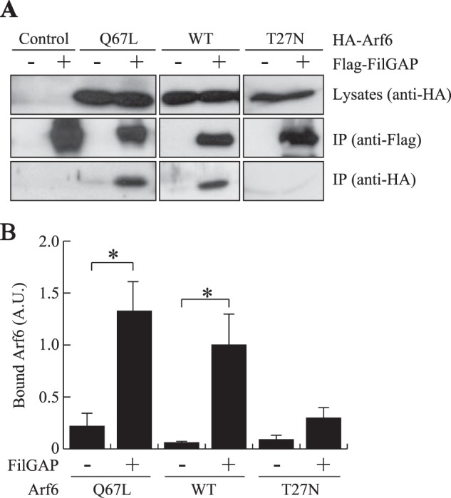
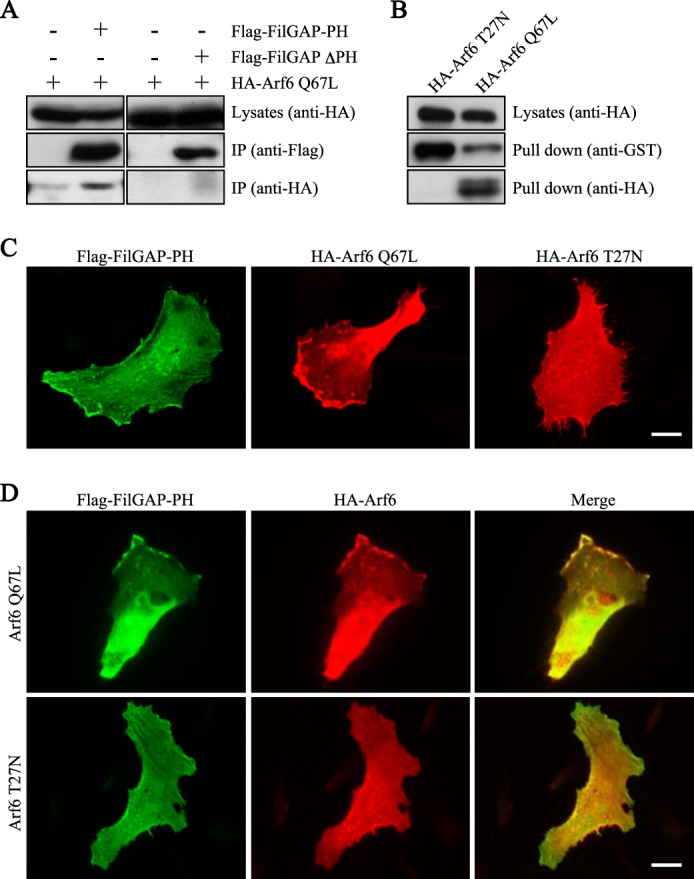
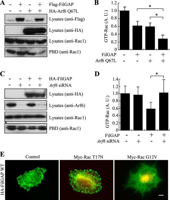
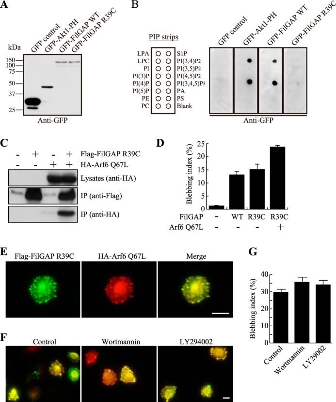
The small GTP-binding protein Arf6 reorganizes the actin cytoskeleton through the regulation of Rac activity. We identified FilGAP, a Rac-specific Rho GTPase-activating protein that is recruited to plasma membranes by binding to activated Arf6. FilGAP binds to Arf6 through its pleckstrin homology domain. Activated Arf6 stimulated RacGAP activity of FilGAP, and knockdown of endogenous Arf6 by siRNA suppresses FilGAP-mediated bleb formation. Mutant FilGAP lacking phosphatidylinositol 3,4,5-trisphosphate (PIP3) binding (FilGAP R39C) binds to activated Arf6 and induces bleb formation. Moreover, bleb formation induced by wild-type FilGAP occurs in the presence of phosphatidylinositol 3-kinase inhibitors, suggesting a PIP3-independent interaction between FilGAP and Arf6. We propose that FilGAP may function as a mediator of the regulation of Rac by Arf6.

Keywords: Actin; Arf6; Cell Migration; Cell Polarization; Cytoskeleton; PI 3-kinase; Rac; Rho; Signal Transduction; Small GTPases.


Figures

FIGURE 1.
FIGURE 1.
The PH domain of FilGAP is required for bleb formation. A, schematic of mutated FilGAP constructs. CC, coiled coil. B, formation of membrane blebbing induced by FilGAP and its mutants. A7 cells were transfected with HA-tagged FilGAP or its mutants for 24 h in growing medium. The cells were fixed and stained with anti-HA antibody. Scale bar = 10 μm. C, sequence alignment of the PH domains. Identical amino acids are shown as boldface letters. FilGAP has a di-Gly, high-affinity, selective binding site to PIP3, as indicated by red letters. Sequence alignment was performed with the assistance of Higgins multiple alignment programs.
FIGURE 2.
FIGURE 2.
Binding of FilGAP to Arf6. A, association of FilGAP and Arf6 in intact cells. HEK cells were transiently transfected with FLAG-tagged FilGAP and HA-tagged Arf6. Cell extracts were prepared, and FLAG-FilGAP was immunoprecipitated (IP) using an anti-FLAG antibody. The washed immunoprecipitates were immunoblotted for the presence of FilGAP and Arf6. B, the relative amount of bound Arf6 proteins, treated as in A, was calculated, and the data are expressed as the mean ± S.E. (n = 4). *, p < 0.05. Statistical significance was determined by Student's t test. A. U., arbitrary units.
FIGURE 3.
FIGURE 3.
The PH domain of FilGAP interacts with Arf6. A, association of the PH domain of FilGAP and Arf6 in intact cells. HEK cells were transiently transfected with HA-Arf6 Q67L and the FLAG-tagged PH domain of FilGAP (FilGAP-PH) or the FLAG-tagged FilGAP mutant lacking the PH domain (FLAG-FilGAP-ΔPH). Cell extracts were prepared, and FilGAP-PH or FLAG-FilGAP-ΔPH protein was immunoprecipitated (IP) using an anti-FLAG antibody. The washed immunoprecipitates were immunoblotted for the presence of the PH domain or FilGAP-ΔPH (anti-FLAG) and Arf6 (anti-HA). B, association of FilGAP and Arf6 in vitro. HEK cells were transiently transfected with HA-Arf6 Q67L or HA-Arf6 T27N. Cell extracts were prepared and incubated with GST-FilGAP-PH fusion protein coupled to glutathione-Sepharose beads. The beads were washed, and the precipitates were immunoblotted for the presence of GST-FilGAP-PH (anti-GST) and Arf6 (anti-HA). C, localization of the PH domain of FilGAP and Arf6 mutants in migrating HeLa cells. HeLa cells were fixed 12 h after transfection with the PH domain of FilGAP or Arf6 mutants. The PH domains of FilGAP and Arf6 mutants were localized by staining the cells with polyclonal anti-FLAG antibody for FilGAP-PH (green) and monoclonal anti-HA antibody for Arf6 Q67L and Arf6 T27N (red). Scale bar =10 μm. D, HeLa cells were fixed 12 h after cotransfection with FLAG-FilGAP-PH and HA-Arf6 Q67L or HA-Arf6 T27N. The PH domains of FilGAP and Arf6 mutants were localized by staining the cells with polyclonal anti-FLAG antibody for FilGAP-PH (green) or monoclonal anti-HA antibody for Arf6 Q67L (red). Merged fluorescent images are also shown. Scale bar = 10 μm.
FIGURE 4.
FIGURE 4.
Involvement of Arf6 in bleb formation induced by FilGAP. A, localization of Arf6 in A7 cells. Human melanoma A7 cells were transfected with HA-tagged, constitutively activated Arf6 Q67L or dominant-negative Arf6 T27N. After 24 h, the cells were fixed, and Arf6 proteins were localized by staining the cells with anti-HA antibody (green). F-actin was localized by TRITC-phalloidin (red). Merged fluorescent images are also shown. Scale bar = 10 μm. B, the percentage of cells showing membrane-associated Arf6 proteins, as shown in A, was calculated, and the data are expressed as the mean ± S.E. (n = 4). *, p < 0.001. Statistical significance was determined by Student's t test. C, immunoblot analysis showing that Arf6 is depleted after 24 h of siRNA treatment of A7 cells. Anti-tubulin antibody was used as a loading control. D, reduction of bleb formation in cells treated with Arf6 siRNA. A7 cells were treated with or without Arf6 siRNA in the presence of an HA-tagged FilGAP construct. After 24 h, the cells were fixed, and FilGAP was stained with anti-HA antibody (green) and TRITC-labeled phalloidin for F-actin (red). Representative merged images are shown. Scale bar = 10 μm. E, the percentage of bleb-positive cells, as shown in D, was calculated, and the data are expressed as the mean ± S.E. (n = 4). *, p < 0.001. Statistical significance was determined by Student's t test. F, requirement of the PH and GAP domains of FilGAP for Arf6-induced bleb formation. A7 cells were transfected with HA-Arf6 Q67L and FLAG-tagged FilGAP or its mutants. After 24 h, the cells were fixed, and FilGAP was stained with rabbit polyclonal anti-FilGAP antibody (green), and HA-Arf6 Q67L was stained with mouse monoclonal anti-HA antibody (red). Representative merged images are shown. Scale bar = 5 μm.
FIGURE 5.
FIGURE 5.
GTPase-stimulating activity of FilGAP and its regulation by Arf6. A, HEK cells were transfected with HA-Arf6 Q67L or FLAG-FilGAP. Cell extracts were incubated with GST-PBD that was immobilized on glutathione-Sepharose beads. The amount of Rac1 in cell lysates before pulldown and GTP (GST-PBD-bound) Rac1 was detected by immunoblotting. Expression levels of transfected Arf6 Q67L (anti-HA) and FilGAP (anti-FLAG) are also shown. B, the relative amount of GTP-Rac1 treated in A was calculated and expressed as the mean ± S.E. (n = 6). *, p < 0.05. Statistical significance was determined by Student's t test. A. U., arbitrary units. C, HEK cells were treated with or without Arf6 siRNA in the presence or absence of the HA-FilGAP construct. Cell extracts were incubated with GST-PBD that was immobilized on glutathione-Sepharose beads. The amount of Rac1 in cell lysates before pulldown and GTP (GST-PBD bound) Rac1 was detected by immunoblotting. Expression levels of transfected HA-FilGAP (anti-HA) and endogenous Arf6 (anti-Arf6) are also shown. The asterisk indicates a nonspecific band. D, the relative amount of GTP-Rac1 treated in C was calculated and expressed as the mean ± S.E. (n = 6). *, p < 0.05. Statistical significance was determined by Student's t test. E, FilGAP induces blebbing through inactivation of Rac1. A7 cells were transfected with HA-FilGAP in the presence or absence of dominant-negative Rac1 (myc-Rac T17N) or constitutively activated Rac1 (myc-Rac G12V) mutants. After 24 h, the cells were fixed, and FilGAP was stained with rabbit polyclonal anti-FilGAP antibody (green) and mouse monoclonal anti-myc antibody for myc-Rac1 mutants (red). Representative merged images of cells are shown. Scale bar = 5 μm.
FIGURE 6.
FIGURE 6.
Arf6 interacts with FilGAP in a PIP3-independent manner. A, HEK cells were transiently transfected with a control plasmid (pEGFP), a pAcGFP plasmid encoding the PH domain of Akt1, or a pEGFP plasmids encoding FilGAP WT or FilGAP R39C. Cell lysates were prepared and immunoblotted with anti-GFP antibody. B, the FilGAP R39C mutant does not bind to PIP3. PIP strips were incubated with the cell lysates in A, and strip-bound proteins were detected using anti-GFP antibody. LPA, lysophosphatidic acid; LPC, lysophosphatidylcholine; PE, phosphatidylethanolamine; PC, phosphatidylcholine; S1P, sphingosine-1-phosphate; PA, phosphatidic acid; PS, phosphatidylserine. C, association of mutant FilGAP R39C and Arf6 in intact cells. HEK cells were transiently transfected with FLAG-FilGAP R39C and HA-Arf6 Q67L. Cell extracts were prepared, and FLAG-FilGAP R39C was immunoprecipitated (IP) using an anti-FLAG antibody. The washed immunoprecipitates were immunoblotted for the presence of FilGAP R39C (anti-FLAG) and Arf6 Q67L (anti-HA). D, bleb formation in cells transfected with FilGAP R39C. A7 cells were transfected with FLAG-FilGAP constructs and an HA-Arf6 Q67L construct. After 24 h, the cells were fixed, and FilGAP and Arf6 Q67L were stained with anti-FLAG (FilGAP) anti-HA (Arf6) antibodies. The percentage of bleb-positive cells was calculated, and the data are expressed as the mean ± S.E. (n = 4). E, localization of Arf6 and FilGAP R39C in A7 cells. A7 cells were transfected with FLAG-tagged mutant FilGAP R39C and constitutively activated Arf6 Q67L. After 24 h, the cells were fixed, and FilGAP R39C (green) and Arf6 Q67L proteins (red) were localized by staining the cells with anti-FLAG and anti-HA antibodies. A merged image is also shown. Scale bar = 10 μm. F, Arf6 stimulates FilGAP activity in a PIP3-independent manner. A7 cells were transfected with wild-type FLAG-FilGAP in the presence of HA-Arf6 Q67L. After 24 h, the cells were incubated with or without of wortmannin (100 nm) or LY294002 (50 μm) for 1 h. The cells were then fixed and stained for FilGAP (green) and Arf6 (red) using anti-FLAG and anti-HA antibodies. Representative merged images are shown. Scale bar = 10 μm. G, the percentage of bleb-positive cells was calculated, and the data are expressed as the mean ± S.E. (n = 4).

References

    1. Jaffe A. B., Hall A. (2005) Rho GTPases. Biochemistry and biology. Annu. Rev. Cell Dev. Biol. 21, 247–269 - PubMed
    1. Donaldson J. G., Honda A. (2005) Localization and function of Arf family GTPases. Biochem. Soc. Trans. 33, 639–642 - PubMed
    1. D'Souza-Schorey C., Chavrier P. (2006) ARF proteins. Roles in membrane traffic and beyond. Nat. Rev. Mol. Cell Biol. 7, 347–358 - PubMed
    1. Myers K. R., Casanova J. E. (2008) Regulation of actin cytoskeleton dynamics by Arf-family GTPases. Trends Cell Biol. 18, 184–192 - PMC - PubMed
    1. Schweitzer J. K., Sedgwick A. E., D'Souza-Schorey C. (2011) ARF6-mediated endocytic recycling impacts cell movement, cell division and lipid homeostasis. Semin. Cell Dev. Biol. 22, 39–47 - PMC - PubMed

Publication types

MeSH terms

Substances