. 2014 Apr 1;20(7):1768-78.
doi: 10.1158/1078-0432.CCR-13-2697. Epub 2014 Feb 13.

T-cell responses to oncogenic merkel cell polyomavirus proteins distinguish patients with merkel cell carcinoma from healthy donors

Affiliations

T-cell responses to oncogenic merkel cell polyomavirus proteins distinguish patients with merkel cell carcinoma from healthy donors

Rikke Lyngaa et al. Clin Cancer Res. .

Abstract

Purpose: Merkel cell carcinoma (MCC) is a highly aggressive skin cancer with strong evidence of viral carcinogenesis. The association of MCC with the Merkel cell polyomavirus (MCPyV) may explain the explicit immunogenicity of MCC. Indeed, MCPyV-encoded proteins are likely targets for cytotoxic immune responses to MCC as they are both foreign to the host and necessary to maintain the oncogenic phenotype. However, to date only a single MCPyV-derived CD8 T-cell epitope has been described, thus impeding specific monitoring of T-cell responses to MCC.

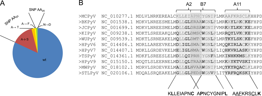
Method: To overcome this limitation, we scanned the MCPyV oncoprotein large T and small T antigens and the virus capsid protein VP1 for potential T-cell epitopes, and tested for MHC class I affinity. We confirmed the relevance of these epitopes using a high-throughput platform for T-cell enrichment and combinatorial encoding of MHC class I multimers.

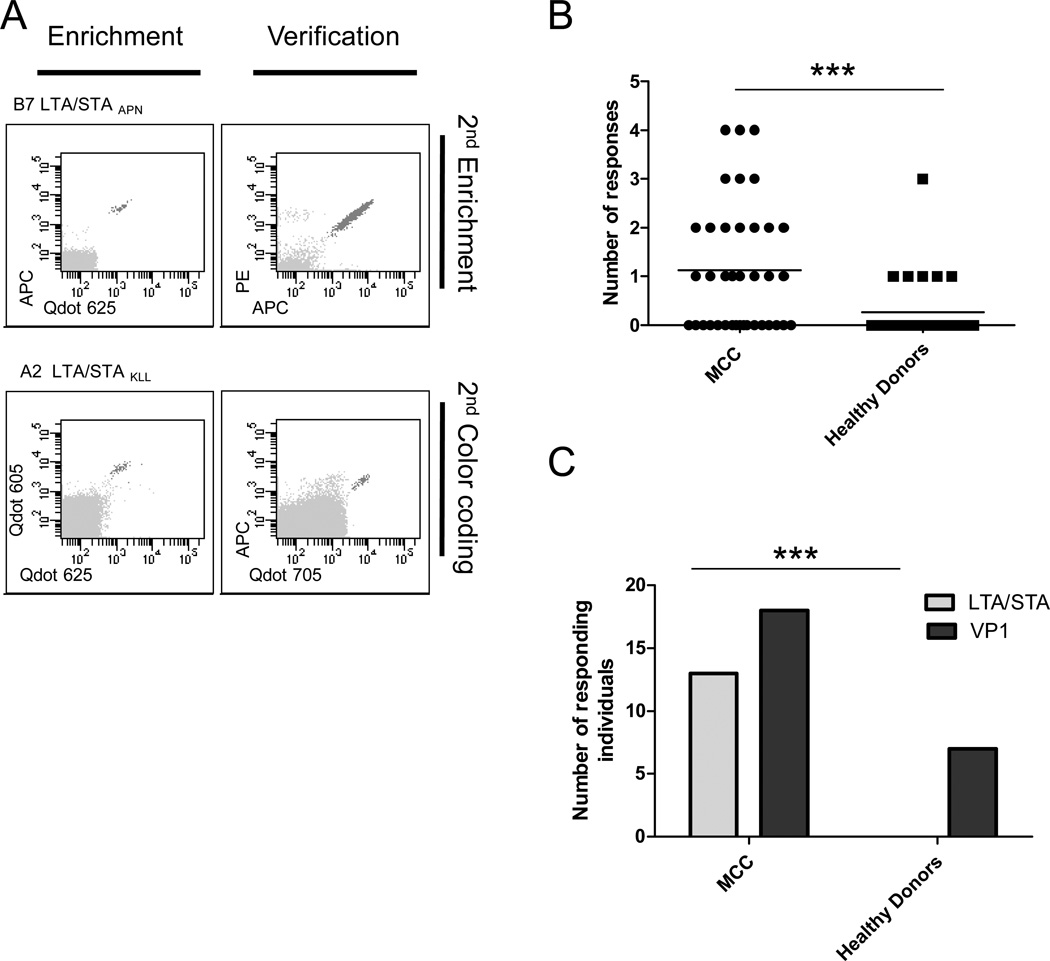
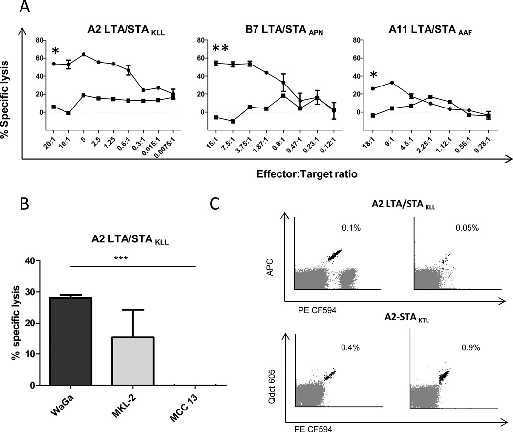
Results: In peripheral blood from 38 patients with MCC and 30 healthy donors, we identified 53 MCPyV-directed CD8 T-cell responses against 35 different peptide sequences. Strikingly, T-cell responses against oncoproteins were exclusively present in patients with MCC, but not in healthy donors. We further demonstrate both the processing and presentation of the oncoprotein-derived epitopes, as well as the lytic activity of oncoprotein-specific T cells toward MHC-matched MCC cells. Demonstrating the presence of oncoprotein-specific T cells among tumor-infiltrating lymphocytes further substantiated the relevance of the identified epitopes.

Conclusion: These T-cell epitopes represent ideal targets for antigen-specific immune therapy of MCC, and enable tracking and characterization of MCPyV-specific immune responses.


Conflict of interest statement

The authors declare no conflicts of interest

Figures

Fig. 1
Fig. 1
T-cell responses against MCPyV in MCC patients and healthy donors. (A) After T-cell enrichment, specific responses were detected using flow cytometry and combinatorial encoded MHC- multimers followed by verification with either a 2nd enrichment (top) or a 2nd MHC-multimer detection using a new color code (bottom). Representative examples from two MCC patients showing LTA/STA directed T-cell responses: MCC#27 B7-LTA/STAAPN (top row) and MCC#8 A2-LTA/STAKLL (bottom row). (B) The number of MCPyV specific T-cell responses per individual in MCC patients and healthy donors, every dot represents one individual. Asterisks indicate significant levels, two-tailed unpaired T-test. (C) The number of MCC patients or healthy donors where LTA/STA or VP1 specific T-cell responses were found. Asterisks indicate significant levels, Fisher`s exact test.
Fig. 2
Fig. 2
Processing and presentation of MCPyV LTA allows T-cell mediated killing by MCPyV-specific T cells. (A) T-cell mediated killing of HLA-expressing LTA transfected K562 cells (Fig. S4A) detected by a flow cytometry-based cytotoxicity assay. The Y-axis shows percent specific lysis at different effector:target ratios with T cells recognizing A2-LTA/STAKLL (left), B7-LTA/STAAPN (middle), and A11-LTS/STAAAF (right). Reactive T-cell populations were enriched to purities ranging from 92–95% of CD8 T cells. Circles: K562 HLA+ LTA were stained with CSFE and K562 HLA were stained with FR. Squares: Unspecific lysis of HLA mis-matched K562 cells stained with CSFE and a second HLA mis-matched K562 was stained with FR for identifying the background lysis. Specific lysis was measured and calculated as described in material and methods. Each point represent the mean of two replicates and error bars the SEM, n=1. Asterisk indicates significant levels. one-tailed unpaired T-test. (B) Chromium release assay was performed using the 51Cr-labeled target cells: MCPyV+/HLA-A2+ MCC cell line WaGa preincubated with INFγ (Supplementary Fig. S4B), MCPyV+/HLA-A2+ MCC cell line MKL-2, and the MCPyV-negative A2+ MCC cell line MCC 13. Target cells were incubated at effector/target cell ratio of 1:1 in duplicates for 4 h before 51Cr release was measured. Bars represent the mean of two replicates and error bars the SEM, n=1. Asterisk indicates significant levels, two-tailed unpaired T-test. (C) Dot plot showing A2-LTAKLL or A2-STAKTL specific CD8 T cells as detected in TIL cultures from a total of 4 MCC patients with MHC mulitmers (APC/PE-CF594 for A2-LTA/STAKLL and Qdot605/ PE-CF594 for A2-STAKTL). The frequency of LTA/STA specific T cells in CD8 T cells is given.
Fig. 3
Fig. 3
Sequence homology of MCPyV-derived CD8 T-cell epitopes within different strains of MCPyV and among other human polyomaviruses. (A) Circle diagram illustrating the variation within the T-cell epitope sequences of LTA from 88 different strains of MCPyV. 17% of the sequences carried a SNP at amino acid position 20, located in both the A2-LTAKLL and B7-LTAAPN epitope causing the indicated changes in the amino acid sequence. (B) Alignment of the MCPyV LTA amino-acid sequence (top) with the corresponding amino-acid sequences from the 10 other known human polyomaviruses, BKPyV, JCPyV, KIPyV, WUPyV, HPyV6, HPyV7, HPyV8, HPyV9, MWPyV and STLPyV. Grey boxes represent the position of the MCPyV-derived A2-, A11-, and B7-restricted epitopes. Amino acids from the other human polyomaviruses that differ from the MCPyV sequence are highlighted in bold. Below, in each epitope the anchor residues are in bold and the auxiliary anchor residues are underlined.

References

    1. Lemos BD, Storer BE, Iyer JG, Phillips JL, Bichakjian CK, Fang LC, et al. Pathologic nodal evaluation improves prognostic accuracy in Merkel cell carcinoma: analysis of 5823 cases as the basis of the first consensus staging system. J Am Acad Dermatol. 2010;63:751–761. - PMC - PubMed
    1. Friedlaender MM, Rubinger D, Rosenbaum E, Amir G, Siguencia E. Temporary regression of Merkel cell carcinoma metastases after cessation of cyclosporine. Transplantation. 2002;73:1849–1850. - PubMed
    1. Heath M, Jaimes N, Lemos B, Mostaghimi A, Wang LC, Peñas PF, et al. Clinical characteristics of Merkel cell carcinoma at diagnosis in 195 patients: the AEIOU features. J Am Acad Dermatol. 2008;58:375–381. - PMC - PubMed
    1. Paulson KG, Iyer JG, Tegeder AR, Thibodeau R, Schelter J, Koba S, et al. Transcriptome-wide studies of merkel cell carcinoma and validation of intratumoral CD8+ lymphocyte invasion as an independent predictor of survival. J Clin Oncol. 2011;29:1539–1546. - PMC - PubMed
    1. Rodig SJ, Cheng J, Wardzala J, Dorosario A, Scanlon JJ, Laga AC, et al. Improved detection suggests all Merkel cell carcinomas harbor Merkel polyomavirus. J Clin Invest. 2012;122:4645–4653. - PMC - PubMed

Publication types

MeSH terms