. 2015 Dec;30(8):738-45.
doi: 10.1177/1533317513520214. Epub 2014 Feb 13.

Detection of Amyloid β Signature in the Lens and Its Correlation in the Brain to Aid in the Diagnosis of Alzheimer's Disease

Affiliations

Detection of Amyloid β Signature in the Lens and Its Correlation in the Brain to Aid in the Diagnosis of Alzheimer's Disease

Charles Kerbage et al. Am J Alzheimers Dis Other Demen. 2015 Dec.

Abstract

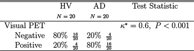
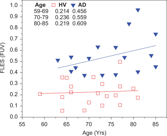
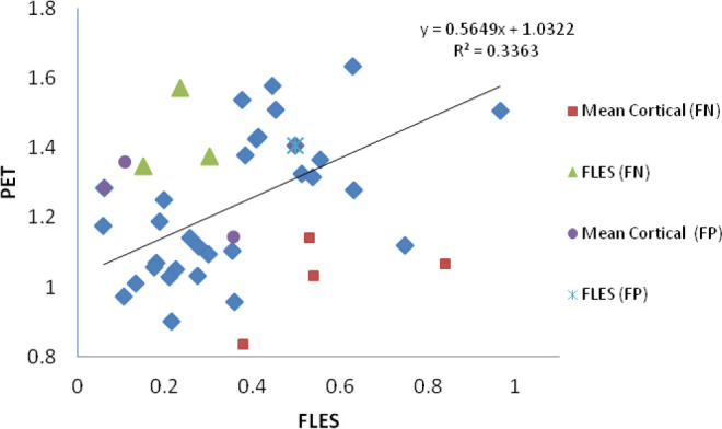
We report the findings from a clinical trial in which a group of patients clinically diagnosed with probable Alzheimer's disease (AD) were discriminated from an age-matched group of healthy volunteers (HVs) with statistical significance (P<.001). The results from 20 patients with AD and 20 HVs were obtained by a Fluorescent Ligand Eye Scanning (FLES) technique that measures a fluorescent signature specific to an exogenous ligand bound to amyloid-β in the lens of the eye. Sensitivity and specificity of 85% and 95%, respectively, have been achieved in predicting clinical diagnosis. Additionally, amyloid brain imaging using florbetapir F18 positron emission tomography shows significant correlation with the results obtained in the eye. Results of the study demonstrate the safety of the FLES system.

Keywords: Alzheimer’s disease; amyloid PET imaging; amyloid-β; fluorescence spectroscopy; human lens.


Conflict of interest statement

The authors declared the following potential conflicts of interest with respect to the research, authorship, and/or publication of this article: Dr Sadowsky, Dr Tariot, Dr Agronin and Dr Alva were principal investigators on the study. None of the investigators have a conflict of interest with the sponsor. Dr.Turner, the study statistician, is an independent contractor to Cognoptix, Inc.

Figures

Figure 1.
Figure 1.
Time and event schedule. Partial exemption allowed inclusion of this test performed within 12 months. Vital signs were taken within 7 minutes of each administration of the ointment. Patients were seated and nonactive for 5 minutes.
Figure 2.
Figure 2.
Demographics of all patients in the study.
Figure 3.
Figure 3.
Scatter plot of the FUV for all patients in the study. The patients are grouped as clinically diagnosed as HV or with AD. The values differed significantly among diagnostic groups (85% sensitivity, 95% specificity, P < .0001, 95% CI, threshold = 0.36). The color coded represents the diagnosis by PET read as positive (red) or negative (black) for AD. FUV indicates fluorescence uptake value; HV, healthy volunteer; AD, Alzheimer’s disease; Ci, confidence interval; PET, positron emission tomography.
Figure 4.
Figure 4.
Efficacy results of FLES to predict clinical diagnosis. FLES indicates Fluorescent Ligand Eye Scanning.
Figure 5.
Figure 5.
Efficacy results of Amyvid PET to predict clinical diagnosis. PET indicates positron emission tomography.
Figure 6.
Figure 6.
Fluorescent Ligand Eye Scanning correlation with Amyvid PET by clinical diagnosis.
Figure 7.
Figure 7.
Fluorescence uptake value assessment as a function of age in both the group with AD and the HV group. The HV group did not exhibit a substantial increase in the FUV with age while a significant increase in FUV was observed in the AD group. The age at which both groups can be discriminated is 55 years old. FUV indicates fluorescence uptake value; HV, healthy volunteer; AD, Alzheimer’s disease.
Figure 8.
Figure 8.
Positron emission tomography (mean cortical) and FLES (FUV) results obtained for all patients. There is a linear regression correlation (r2 = .33, P < .0006) between both methods that correctly predict clinical diagnosis. Overall, FLES did not correctly predict 4 patients; 3 false negatives (FN) and 1 false positive (FP). Visual PET did not correctly predict 8 patients; 4 FNs and 4 FPs. FLES indicates Fluorescent Ligand Eye Scanning; PET, positron emission tomography; FUV, fluorescence uptake value.

References

    1. Hebert LE, Weuve J, Scherr PA, Evans DA. Alzheimer disease in the United States (2010-2050) estimated using 2010 census. Neurology. 2013;80(19):1778–1783. - PMC - PubMed
    1. Thies W, Bleiler L; Alzheimer's Association. 2013. Alzheimer’s Disease Facts and Figures. Alzheimers Dement. 2013;9(2):208–245. - PubMed
    1. Hyman BT, Phelps CH, Beach TG, et al. . National institute on aging-Alzheimer’s association guidelines for the neuropathologic assessment of Alzheimer’s disease. Alzheimers Dement. 2012;8(1):1–13. - PMC - PubMed
    1. McKhann GM, Knopman DS, Chertkow H, et al. . The diagnosis of dementia due to Alzheimer’s disease: recommendations from the national institute of aging-Alzheimer’s association workgroups on diagnostic guidelines for Alzheimer’s disease. Alzheimers Dement. 2011;7(3):263–269. - PMC - PubMed
    1. McKhann G, Drachman D, Folstein M, Katzman R, Price D, Stadlan EM. Clinical diagnosis of Alzheimer's disease: report of the NINCDS-ADRDA work group under the auspices of department of health and human services task force on Alzheimer's disease. Neurology. 1984;34(7):939–944. - PubMed

Publication types

LinkOut - more resources