Huge giant cell tumor of the sacrum: A case report
- PMID: 24527097
- PMCID: PMC3919884
- DOI: 10.3892/ol.2014.1812
Huge giant cell tumor of the sacrum: A case report
Abstract
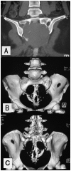
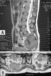
The current report describes the case of a 29-year-old female with a sacral giant cell tumor (GCT) during pregnancy. Originally, the patient presented with severe pain in the lumbosacral region, radiating posterolaterally from the lumbar spine into the bilateral thigh and subsequently, into the bilateral crus posterolaterally. Plain X-rays, computed tomography and magnetic resonance imaging showed osteolytic destruction of the sacrococcygeal bones and a huge soft-tissue mass with features of a chordoma. The patient underwent a partial en bloc sacrectomy (partial S1 and completely below) and curettage for tumors located at the sacroiliac joint and underlying left ilium, with bilateral internal iliac arteries ligated to control intraoperative hemorrhage. The patient's bilateral S2 nerve roots were killed. The diagnosis of conventional GCT was determined based on the histopathological examination of the resected specimen. Urinary and bowel functions were recovered by exercising.
Keywords: S2 nerve root; chordoma; giant cell tumor; rectum; sacral tumor; urinary and bowel function.
Figures


References
-
- Bloem JL, Reidsma II. Bone and soft tissue tumors of hip and pelvis. Eur J Radiol. 2012;81:3793–3801. - PubMed
-
- Turcotte RE, Sim FH, Unni KK. Giant cell tumor of the sacrum. Clin Orthop Relat Res. 1993;291:215–221. - PubMed
-
- Cowan RW, Singh G, Ghert M. PTHrP increases RANKL expression by stromal cells from giant cell tumor of bone. J Orthop Res. 2012;30:877–884. - PubMed
-
- Leggon RE, Zlotecki R, Reith J, Scarborough MT. Giant cell tumor of the pelvis and sacrum: 17 cases and analysis of the literature. Clin Orthop Relat Res. 2004;423:196–207. - PubMed
-
- Lin PP, Guzel VB, Moura MF, Wallace S, Benjamin RS, Weber KL, et al. Long-term follow-up of patients with giant cell tumor of the sacrum treated with selective arterial embolization. Cancer. 2002;95:1317–1325. - PubMed
LinkOut - more resources
Full Text Sources
Other Literature Sources
Research Materials
