

Migration and coiling of peritoneal catheter into the subgaleal space: a very rare complication of subgaleoperitoneal shunt
- PMID: 24527199
- PMCID: PMC3921284
- DOI: 10.3340/jkns.2013.54.6.525
Migration and coiling of peritoneal catheter into the subgaleal space: a very rare complication of subgaleoperitoneal shunt
Abstract
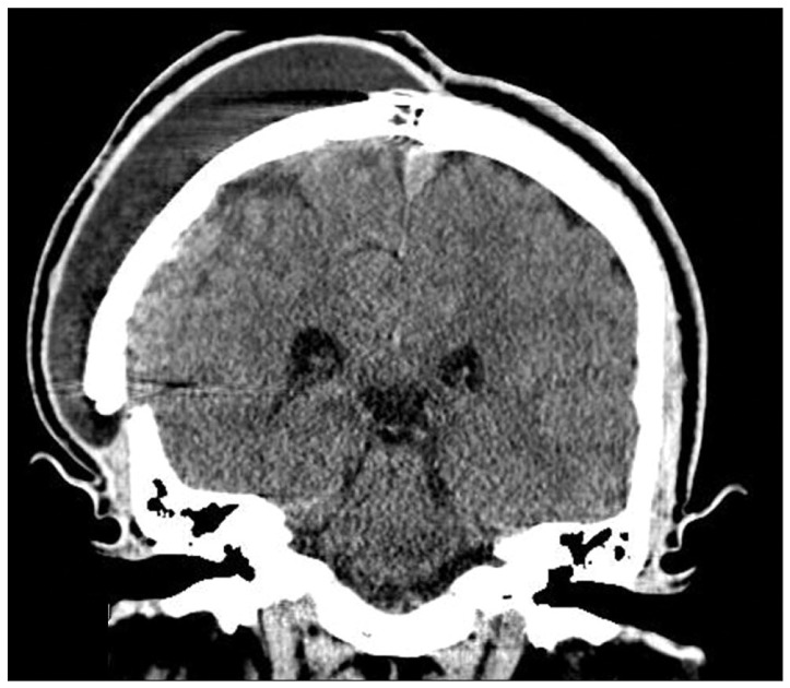
Upward migration of the peritoneal catheter of a subgaleo-peritoneal (SP) shunt and coiling into the subgaleal space is an extremely rare complication of a SP shunt. A 32-year-old male patient visited our hospital presenting with a large skull defect due to a prior craniectomy performed elsewhere. The patient underwent a cranioplasty with methylmetacrylate, but subsequently developed progressive pseudomeningocele and subgaleal cerebrospinal fluid (CSF) collection. The patient underwent CSF diversion via a SP shunt. After SP shunting, the pseudomeningocele disappeared completely. Six months later, the patient presented with progressive scalp swelling. Skull X-ray showed migration and coiling of the distal catheter of the SP shunt. The patient was treated by removing the entire shunt catheter and the dura was covered with a subgaleal flap. We would like to report our experience with a very rare complication of subgaleo-peritoneal shunting.
Keywords: Migration; Pseudomeningocele; Subgaleo-peritoneal shunt.
Figures


Similar articles
-
Distal ventriculoperitoneal shunt catheter tightly coiled around the valve in the absence of a subgaleal cerebrospinal fluid collection: illustrative case.J Neurosurg Case Lessons. 2021 May 17;1(20):CASE21115. doi: 10.3171/CASE21115. eCollection 2021 May 17. J Neurosurg Case Lessons. 2021. PMID: 35855019 Free PMC article.
-
Distal subgaleal-peritoneal shunt migration into the abdominal wall with subsequent formation of a pre-peritoneal pseudocyst: a rare complication.J Surg Case Rep. 2010 Sep 1;2010(7):9. doi: 10.1093/jscr/2010.7.9. J Surg Case Rep. 2010. PMID: 24946343 Free PMC article.
-
Ventriculoperitoneal shunt migration and coiling: A report of two cases.J Pediatr Neurosci. 2012 May;7(2):114-6. doi: 10.4103/1817-1745.102572. J Pediatr Neurosci. 2012. PMID: 23248689 Free PMC article.
-
CSF hydrothorax: neither migration of peritoneal catheter into the chest nor ascites. Case report and review of the literature.Childs Nerv Syst. 2012 Nov;28(11):1843-8. doi: 10.1007/s00381-012-1862-1. Epub 2012 Jul 24. Childs Nerv Syst. 2012. PMID: 22825420 Review.
-
Ventriculosubgaleal shunting-a comprehensive review and over two-decade surgical experience.Childs Nerv Syst. 2018 Sep;34(9):1639-1642. doi: 10.1007/s00381-018-3887-6. Epub 2018 Jul 12. Childs Nerv Syst. 2018. PMID: 30003327 Review.
Cited by
-
Upward Migration and Coiling of the Distal Catheter Toward the Valve Site.Cureus. 2021 Sep 15;13(9):e17993. doi: 10.7759/cureus.17993. eCollection 2021 Sep. Cureus. 2021. PMID: 34667670 Free PMC article.
-
Retrograde Partial Migration of Ventriculoperitoneal Shunt with Chamber: Review of Causative Factors and Its Prevention.J Pediatr Neurosci. 2017 Jan-Mar;12(1):93-95. doi: 10.4103/1817-1745.205654. J Pediatr Neurosci. 2017. PMID: 28553395 Free PMC article.
-
Successful Management of Iatrogenic Cranial Pseudomeningocele With Subgaleal Shunt.Cureus. 2023 Feb 1;15(2):e34513. doi: 10.7759/cureus.34513. eCollection 2023 Feb. Cureus. 2023. PMID: 36874315 Free PMC article.
References
-
- Acharya R, Bhutani A, Saxena H, Madan VS. Complete migration of ventriculoperitoneal shunt into the ventricle. Neurol Sci. 2002;23:75–77. - PubMed
-
- Jang HD, Kim MS, Lee NH, Kim SH. Anal Extrusion of Distal V-P Shunt Catheter after Double Perforation of Large Intestine. J Korean Neurosurg Soc. 2007;42:232–234.
-
- Johnson MC, Maxwell MS. Delayed intrapleural migration of a ventriculoperitoneal shunt. Childs Nerv Syst. 1995;11:348–350. - PubMed
-
- Kiran NA, Thakar S, Mohan D, Aryan S, Rao AS, Hegde AS. Subgaleo-peritoneal shunt: an effective and safer alternative to lumboperitoneal shunt in the management of persistent or recurrent iatrogenic cranial pseudomeningoceles. Neurol India. 2013;61:65–68. - PubMed
Publication types
LinkOut - more resources
Full Text Sources
Other Literature Sources
