

Biomechanics of Scar Tissue and Uninjured Skin
- PMID: 24527323
- PMCID: PMC3840475
- DOI: 10.1089/wound.2011.0321
Biomechanics of Scar Tissue and Uninjured Skin
Abstract
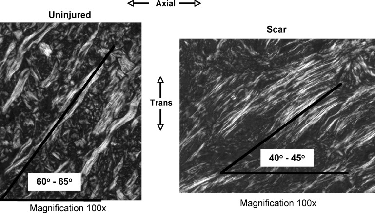
Significance: Skin exhibits direction-dependent biomechanical behavior, influenced by the structural orientation of its collagen-rich fibrous network and its viscous ground-substance matrix. Injury can affect the skin's structure and composition, thereby greatly influencing the biomechanics and directionality of the resulting scar tissue.
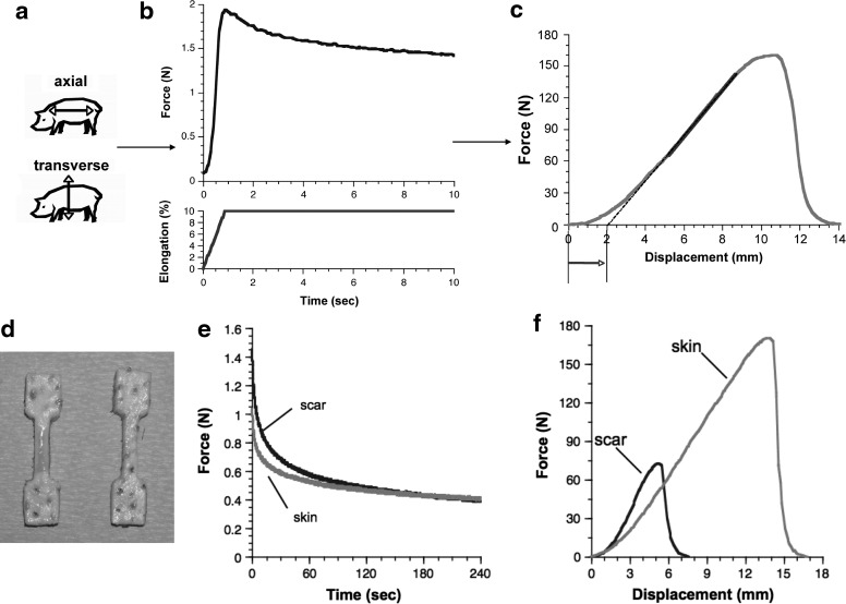
Recent advances: A combination of stress-relaxation and tensile failure testing identifies both the tissue's physiologically relevant viscoelastic behavior and resistance to rupture. When studied in mutually orthogonal directions in the plane of the tissue, these measures give insight into the directional properties of healthy tissue, and how they change with injury. By controlling the biomechanics of the wound environment, a new force-modulating dressing has demonstrated the ability to improve healing and reduce scar formation.
Critical issues: Skin and scar biomechanics are typically characterized by using tensile failure, which identifies the tissue's resistance to rupture but offers limited insight into its normal daily function. Characterizing physiologically relevant biomechanics of skin, and how they change with injury, is critical to understand the tissue's ability to resist elongation, bear load, and dissipate energy via viscous means.
Future directions: Compared with uninjured skin, scar tissue demonstrates similar high-load stiffness, greatly reduced resistance to failure, reduced low-load compliance, and altered material directionality. These findings, identified through combined stress relaxation and failure testing, suggest morphological changes with injury that are consistent with the viscoelastic and directional changes observed biomechanically. A more complete understanding of the directional, physiologically relevant skin biomechanics can guide the design and critical functional assessment of wound treatments, scaffolds, and tissue-engineered skin replacements.
Figures

References
-
- Lanir Y. Skin mechanics. In: Skalak R, editor; Chien S., editor. Handbook of Bioengineering. Dallas, TX: McGraw-Hill; 1987. pp. 11.1–11.25.
-
- Lanir Y. The fibrous structure of the skin, its relation to mechanical behaviour. In: Marks R, editor; Payne PA., editor. Bioengineering and the Skin. Hingham, MA: MTP Press; 1981. pp. 93–96.
-
- Smith LT. Holbrook KA. Byers PH. Structure of the dermal matrix during development and in the adult. J Invest Dermatol. 1982;79(Suppl 1):93S. - PubMed
-
- Stark HL. Directional variations in the extensibility of human skin. Br J Plast Surg. 1977;30:105. - PubMed
-
- Dunn MG. Silver FH. Swann DA. Mechanical analysis of hypertrophic scar tissue: structural basis for apparent increased rigidity. J Invest Dermatol. 1985;84:9. - PubMed
Publication types
LinkOut - more resources
Full Text Sources
Other Literature Sources
