Review
. 2013 Jun;2(5):195-214.
doi: 10.1089/wound.2013.0429.

Dynamics of Transforming Growth Factor Beta Signaling in Wound Healing and Scarring

Affiliations
Review

Dynamics of Transforming Growth Factor Beta Signaling in Wound Healing and Scarring

Kenneth W Finnson et al. Adv Wound Care (New Rochelle). 2013 Jun.

Abstract

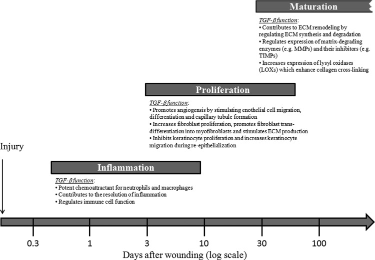
Significance: Wound healing is an intricate biological process in which the skin, or any other tissue, repairs itself after injury. Normal wound healing relies on the appropriate levels of cytokines and growth factors to ensure that cellular responses are mediated in a coordinated manner. Among the many growth factors studied in the context of wound healing, transforming growth factor beta (TGF-β) is thought to have the broadest spectrum of effects.

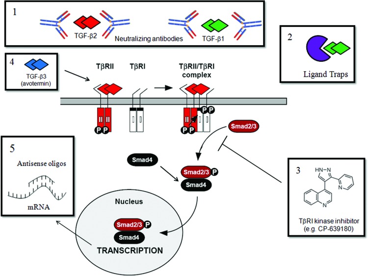
Recent advances: Many of the molecular mechanisms underlying the TGF-β/Smad signaling pathway have been elucidated, and the role of TGF-β in wound healing has been well characterized. Targeting the TGF-β signaling pathway using therapeutic agents to improve wound healing and/or reduce scarring has been successful in pre-clinical studies.

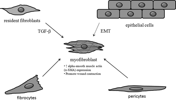
Critical issues: Although TGF-β isoforms (β1, β2, β3) signal through the same cell surface receptors, they display distinct functions during wound healing in vivo through mechanisms that have not been fully elucidated. The challenge of translating preclinical studies targeting the TGF-β signaling pathway to a clinical setting may require more extensive preclinical research using animal models that more closely mimic wound healing and scarring in humans, and taking into account the spatial, temporal, and cell-type-specific aspects of TGF-β isoform expression and function.

Future directions: Understanding the differences in TGF-β isoform signaling at the molecular level and identification of novel components of the TGF-β signaling pathway that critically regulate wound healing may lead to the discovery of potential therapeutic targets for treatment of impaired wound healing and pathological scarring.


Figures

None
Anie Philip, PhD
Figure 1.
Figure 1.
Members of the TGF-β superfamily and their signaling components. TGF-β superfamily members include TGF-β (-β1, -β2, and -β3), activin, nodal, BMPs (-2, -4, and -7), AMH/MIS, and GDF-5. TGF-β superfamily members signal through a unique pair of transmembrane serine-threonine kinases known as the type I and type II receptors to mediate intracellular Smad signaling. The TGF-β/activin/Nodal subfamily binds to ALK 4, 5, and 7 and activates Smads 2 and 3; whereas the BMP/GDF/MIS subfamily generally binds to ALK 1, 2, 3, or 6 and activates Smads 1, 5, and 8. Activated Smad2/3 and Smad1/5/8 form a complex with Smad4 and enter the nucleus, where they regulate target gene expression. Accessory or co-receptors (betaglycan, endoglin, CD109, and cripto) are potent modulators of signaling by TGF-β superfamily members. TGF-β, transforming growth factor beta; BMP, bone morphogenetic protein; AMH, anti-müllerian hormone; MIS, Müllerian inhibiting substance; GDF, growth and differentiation factor; ALK, activin-like receptor kinase. To see this illustration in color, the reader is referred to the web version of this article at www.liebertpub.com/wound
Figure 2.
Figure 2.
Latent and active forms of TGF-β ligand. TGF-β is synthesized as a homo-dimeric proprotein (pro–TGF-β) and undergoes proteolytic cleavage in the trans-Golgi network by furin-like enzymes, giving rise to the mature TGF-β dimer and its pro-peptide, also known as LAP. TGF-β is secreted either as an SLC, which comprises the mature TGF-β dimer in association with LAP, or as an LLC in which the LAP portion of SLC is covalently linked to a protein known as latent TGF-β binding protein (LTBP). (A) SLC: The mature TGF-β dimer (red) is non-covalently associated with its LAP (green). *Asterisks indicate the regions that have undergone proteolytic processing by furin-like enzymes in the trans-Golgi before secretion. (B) LLC: The SLC is covalently linked to latent TGF-β binding protein (LTBP, yellow) by disulfide bonds to form the LLC. (C) Mature TGF-β dimer: The mature TGF-β dimer is released from the latent complex by different mechanisms, giving rise to the active form of TGF-β that can bind its receptors and elicit biological responses. LAP, latency associated peptide; SLC, small latent complex; LLC, large latent complex. To see this illustration in color, the reader is referred to the web version of this article at www.liebertpub.com/wound
Figure 3.
Figure 3.
TGF-β/Smad signal transduction pathway. TGF-β signaling is initiated when the TGF-β ligand binds to the extracellular domain of TβRII. TGF-β1 and TGF-β3 bind TβRII with a high affinity, whereas TGF-β2 requires the TGF-β co-receptor betaglycan (TβRIII) to “present” it to TβRII. TGF-β-associated TβRII then recruits TβRI, resulting in the formation of a heterotetrameric receptor signaling complex comprising one TGF-β ligand, one homo-dimeric TβRII, and one homo-dimeric TβRI. TβRII is a constitutively active kinase that phosphorylates TβRI, resulting in activation of TβRI kinase activity. TβRI then phosphorylates intracellular Smad2 and Smad3 proteins, which, in turn, form a complex with Smad4 and enter the nucleus to regulate gene transcription. TβRII, type II TGF-β receptor. To see this illustration in color, the reader is referred to the web version of this article at www.liebertpub.com/wound
Figure 4.
Figure 4.
Regulation of TGF-β signaling by clathrin-dependent and -independent endocytosis. TGF-β receptors can be internalized by clathrin-dependent and clathrin-independent, membrane-raft dependent mechanisms. TGF-β receptors internalized via clathrin-coated pit mediated endocytosis traffic to Smad anchor for receptor activation (SARA)-containing early endosome and propagate signal transduction. TGF-β receptors in the early endosome can be recycled back to the plasma membrane in a Rab11-dependent manner. TGF-β receptors internalized by membrane-raft dependent endocytosis traffic to caveolin-1 positive vesicles, where they are targeted for Smad7/Smurf2-mediated ubiquitination and proteosomal/lysosomal degradation. Other potential intracellular trafficking pathways for TGF-β receptors, including bi-directional trafficking between the early endosome and caveolin-1 positive vesicles (intermediate pathways) or direct trafficking of TGF-β receptors from the early endosome to proteosomal/lysosomal degradation pathways, are current topics of investigation. Smurf, Smad ubiquitination regulatory factor. To see this illustration in color, the reader is referred to the web version of this article at www.liebertpub.com/wound
Figure 5.
Figure 5.
Schematic diagram showing the role of TGF-β in regulating all three phases of the wound healing process. Inflammation: TGF-β acts as a potent chemoattractant for immune cells (neutrophils and macrophages) during the early stages of inflammation, regulates immune cell function, and contributes to resolution of inflammation. Proliferation: TGF-β promotes angiogenesis by stimulating endothelial cell migration, differentiation, and capillary tubule formation. TGF-β also stimulates fibroblast proliferation, promotes fibroblast trans-differentiation into myofibroblasts, and stimulates ECM production. In addition, TGF-β inhibits keratinocyte proliferation and enhances keratinocyte migration, promoting re-epithelialization. Maturation: TGF-β regulates the balance of ECM synthesis and degradation by tightly controlling the production of ECM components and regulating their rate of degradation by modulating synthesis of MMPs and production of protease inhibitors such as TIMPs. TGF-β also regulates ECM remodeling by stimulating production of LOXs, which play an important role in collagen cross-linking. ECM, extracellular matrix; MMPs, matrix metalloproteinases; TIMPs, tissue inhibitors of matrix metalloproteinases; LOXs, lysyl oxidases.
Figure 6.
Figure 6.
Myofibroblasts originate from different cell types and play a critical role in wound healing and scarring. Myofibroblasts are specialized cells that express alpha smooth muscle actin (α-SMA, red) and display a contractile phenotype. During wound healing, resident fibroblasts trans-differentiate into myofibroblasts in response to TGF-β and promote wound contraction. Fibrocytes are circulating bone marrow-derived cells that can enter tissues and differentiate into myofibroblasts in response to TGF-β. Myofibroblasts also originate from other cell types such as epithelial cells through epithelial-to-mesenchymal transition (EMT) and perivascular cells (pericytes) by trans-differentiation, and these processes have been implicated in the pathogenesis of hypertrophic scarring.
Figure 7.
Figure 7.
Characteristics of human hypertrophic scars and keloids. (A) Hypertrophic scar: This appears as a red, raised scar that does not extend beyond the boundaries of the original injury. They have nodular collagen deposits containing α-SMA producing myofibroblasts that are involved in scar contracture. Hypertrophic scars can regress with time. The main findings from studies on the role of TGF-β signaling and hypertrophic scarring are indicated. (B) Keloid: This appears as a shiny and smooth protuberance ranging from pink to purple in color and extends beyond the boundaries of the original wound. Unlike hypertrophic scars, keloids do not have nodular collagen deposits, α-SMA-producing myofibroblast, do not undergo scar contracture, and do not regress with time. The main findings from studies on the role of TGF-β signaling and keloid formation are indicated. Images were obtained with permission from the DermNet NZ Web site (www.dermnetnz.org). To see this illustration in color, the reader is referred to the web version of this article at www.liebertpub.com/wound
Figure 8.
Figure 8.
Therapeutic strategies that have been used to target the TGF-β signaling pathway to reduce scarring. (1) Neutralizing TGF-β antibodies and (2) ligand traps bind TGF-β and neutralize TGF-β activity by preventing its binding to the TGF-β signaling receptors (TβRII and TβRI). (3) Small molecules that inhibit ALK5 kinase activity, including CP-639180 (depicted), prevent ALK5-induced phosphorylation of Smad2 and Smad3 and downstream signaling events. (4) Recombinant TGF-β3 protein (avotermin) binds to TGF-β signaling receptors and elicits Smad2/3-dependent signaling. Unlike TGF-β1 and TGF-β2 isoforms that have pro-scarring effects, TGF-β3 has anti-scarring properties. The molecular mechanisms underlying the different responses of the TGF-β isoforms have not been elucidated. (5) Antisense oligonucleotides are single-stranded DNA or RNA sequences that are complementary to a specific mRNA sequence. They bind to their target mRNA sequence and silence gene expression by blocking protein translation or promoting degradation of the mRNA transcript. To see this illustration in color, the reader is referred to the web version of this article at www.liebertpub.com/wound

References

    1. Penn JW. Grobbelaar AO. Rolfe KJ. The role of the TGF-β family in wound healing, burns and scarring: a review. Int J Burns Trauma. 2012;2:18. - PMC - PubMed
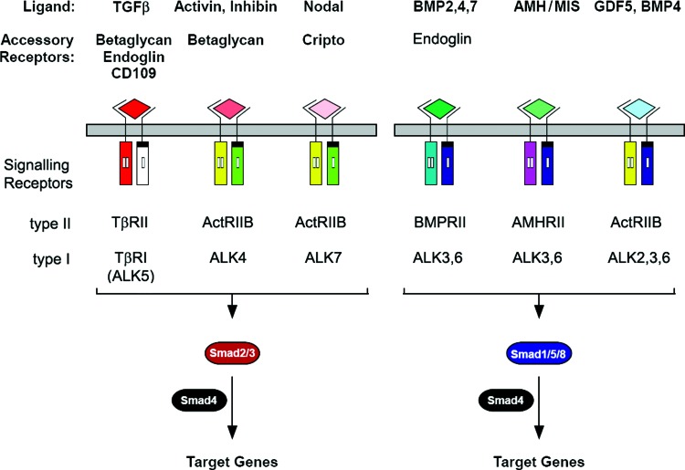
    1. Wu MY. Hill CS. TGF-β superfamily signaling in embryonic development and homeostasis. Dev Cell. 2009;16:329. - PubMed
    1. Biernacka A. Dobaczewski M. Frangogiannis NG. TGF-β signaling in fibrosis. Growth Factors. 2011;29:196. - PMC - PubMed
    1. Rider CC. Mulloy B. Bone morphogenetic protein and growth differentiation factor cytokine families and their protein antagonists. Biochem J. 2010;429:1. - PubMed
    1. Schmierer B. Hill CS. TGF-β/Smad signal transduction: molecular specificity and functional flexibility. Nat Rev Mol Cell Biol. 2007;8:970. - PubMed