Review
. 2013 Jul;2(6):283-295.
doi: 10.1089/wound.2012.0398.

The Role of Endothelial Progenitor Cells in Postnatal Vasculogenesis: Implications for Therapeutic Neovascularization and Wound Healing

Affiliations
Review

The Role of Endothelial Progenitor Cells in Postnatal Vasculogenesis: Implications for Therapeutic Neovascularization and Wound Healing

Swathi Balaji et al. Adv Wound Care (New Rochelle). 2013 Jul.

Abstract

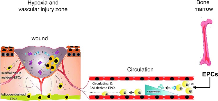
Significance: Postnatal vasculogenesis mediated via endothelial progenitor cells (EPCs) contributes to re-endothelialization and augments neovascularization after ischemia and tissue injury, providing a novel therapeutic application. However, controversy exists with respect to the origin, identification, and contributions of the EPCs to neovascularization, necessitating further study.

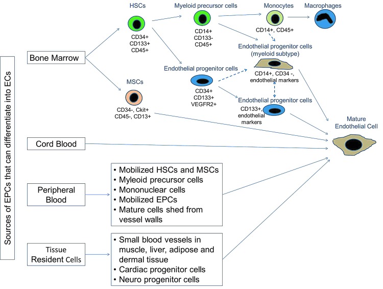
Recent advances: Bone marrow (BM) or circulating cells expressing cd133/vascular endothelial growth factor receptor 2 include those with endothelial progenitor capacity. Increasing evidence suggests that there are additional BM-derived (myeloid; mesenchymal cells) and non-BM-derived (peripheral and cord-blood; tissue-resident) cell populations which also give rise to endothelial cells (ECs) and contribute to re-endothelialization and growth factor release after ischemia and tissue injury. Currently, EPCs are being used as diagnostic markers for the assessment of cardiovascular and tumor risk/progression. Techniques aimed at enhancing ex vivo expansion and the therapeutic potential of these cells are being optimized.

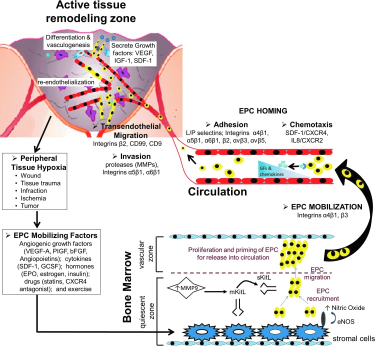
Critical issues: Mobilization and EPC-mediated neovascularization are critically regulated. Stimulatory (growth factors, statins, and exercise) or inhibitory factors (obesity, diabetes, and other cardiovascular diseases) modulate EPC numbers and function. Recruitment and incorporation of EPCs require a coordinated sequence of signaling events, including adhesion, migration (by integrins), and chemoattraction. Finally, EPCs differentiate into ECs and/or secrete angiogenic growth factors. These cells are highly plastic, and depending on the microenvironment and presence of other cells, EPCs transdifferentiate and/or undergo cell fusion and become cells of a different lineage. Therefore, in vitro culture conditions should be optimized to mimic the in vivo milieu to fully characterize the biological function and contribution of EPCs to postnatal vasculogenesis.

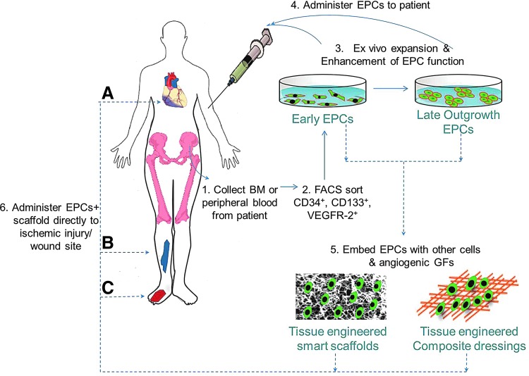
Future directions: Advances in characterization of the EPC biology and enhancement of EPC functions are required. In addition, innovative tissue-engineered carrier matrices that permit embedding of EPCs and provide optimal conditions for EPC survival and endothelial outgrowth will further contribute to EPC-mediated therapeutic applications in wound healing and ischemia repair.


Figures

None
Swathi Balaji, PhD
Figure 1.
Figure 1.
Origin and differentiation of endothelial progenitor cells (EPCs). Several sources have been identified for progenitor cells that can differentiate into endothelial cells. Primarily, hematopoietic stem cells (HSCs) resident in the bone marrow (BM) niche give rise to a population of cells that differentiate into myeloid progenitor or endothelial progenitor subtypes. These subsets differentiate into their respective mature cell type. Alternative pathways (dashed lines) to derive EPCs have been proposed, where EPCs arise directly from HSCs or via a myeloid progenitor/monocyte intermediate. Cell surface markers differentiate cells along the proposed pathway of EPC ontogeny. To see this illustration in color, the reader is referred to the web version of this article at www.liebertpub.com/wound
Figure 2.
Figure 2.
EPCs mobilize in response to hypoxia induced by trauma or vascular injury. In normal homeostatic conditions, EPCs reside within a stem cell niche in the BM. Peripheral tissue hypoxia under trauma or wound healing or vascular injury conditions results in increased production of EPC-mobilizing chemokines and growth factors to a concentration greater than that in the BM, resulting in EPC release and mobilization into the peripheral circulation. Once in circulation, EPCs respond to chemokine signaling in the tissues undergoing active remodeling and home to the injury site. Concomitantly, circulating progenitor cells, tissue-resident, and adipose-derived stem cells respond to the chemokine signaling and home to the active tissue-remodeling site. To see this illustration in color, the reader is referred to the web version of this article at www.liebertpub.com/wound
Figure 3.
Figure 3.
BM-derived EPC stages during adult vasculogenesis—hypoxia/cytokine-driven EPC mobilization from BM to EPC homing and contribution to neovascularization. Circulating EPC count in peripheral blood is very low under normal homeostatic conditions. Hypoxia induced by tissues undergoing active remodeling after injury or ischemia increases the EPC-mobilizing factors, resulting in increase of peripheral blood EPCs. The life cycle of BM-derived EPCs is a complex cascade, which begins with the EPC-mobilizing signals, including angiogenic growth factors, cytokines, hormones, drugs, or exercise, releasing the EPCs from BM. This is mediated via increased matrix metalloproteinase-9 (MMP-9) activity in the quiescent zone of the BM niche, which cleaves the membrane-bound stem cell cytokine mKitL expressed by stromal cells to liberate soluble sKitL. sKitL recruits EPCs and then stimulates them to migrate to the vascular zone of the BM, which is more permissive for proliferation and priming of EPCs for subsequent mobilization into peripheral blood. Additionally, nitric oxide produced by stromal cells is crucial for EPC translocation. Homing and recruitment of EPCs into the active remodeling sites require a coordinated multistep process, including chemoattraction, adhesion, transendothelial migration, and tissue invasion. Locally enhanced levels of cytokines and growth factors provide a chemotaxic trigger for EPCs to home to the sites of remodeling. Recruited EPCs adhere to the endothelium and start transendothelial migration and invasion into the extravascular tissue, where they exert their effects by integration into neovasculature, re-endothelialization, in situ differentiation, and and/or cytokine production. A nonexhaustive list of various cytokines, growth factors, and integrins implicated in each stage of the EPC cycle is illustrated. To see this illustration in color, the reader is referred to the web version of this article at www.liebertpub.com/wound
Figure 4.
Figure 4.
In vitro culture of EPCs: early-versus-late EPCs. Autologous EPCs for transplantation applications are grown from whole peripheral blood mononuclear cells. In vitro culture of these EPCs constitutes two distinct cell populations: early- and late-outgrowth EPCs. The majority of endothelial-like cells interpreted to be EPCs appear to originate from monocytic, CD14-positive cells (early EPCs). There are little data to support their endothelial stem or progenitor status. Alternately, a small population of the early EPCs when cultured for >14 days retains the true progenitor status. These cells are CD14-negative and demonstrate an increased capacity to proliferate and assemble into capillary tubes (late-outgrowth EPCs). Both populations demonstrate an ability to differentiate into endothelial cells and promote neovascularization in vivo, possibly through different mechanisms, that is, supply of angiogenic versus physical integration into newly forming vessels. To see this illustration in color, the reader is referred to the web version of this article at www.liebertpub.com/wound
Figure 5.
Figure 5.
A proposed pathway for EPC contribution to ischemic and cutaneous wound healing and therapeutic angiogenesis. Therapeutic angiogenesis using EPCs can be harnessed by isolating these cells for patients, expanding them in vitro to an appropriate number, and enhancing their therapeutic potential using ex vivo genetic manipulations (gene, liposome, or growth factor therapy) and administering them back to the patient by systemic or local infusion at ischemic (A, B) or wound site (C). Alternately, EPCs can be embedded along with other cells or vulnerary growth factors in innovative tissue-engineered carrier matrices (scaffolds or dressings) that provide physiological, optimal conditions for EPC survival and endothelial outgrowth. Direct application of these EPC-infused scaffolds may further contribute to therapeutic angiogenesis for applications in wound healing and ischemia repair. To see this illustration in color, the reader is referred to the web version of this article at www.liebertpub.com/wound

References

    1. Ko SH. Nauta A. Wong V. Glotzbach J. Gurtner GC. Longaker MT. The role of stem cells in cutaneous wound healing: what do we really know? Plast Reconstr Surg. 2011;127(Suppl 1):10S. - PubMed
    1. Asahara T. Masuda H. Takahashi T. Kalka C. Pastore C. Silver M. Kearne M. Magner M. Isner JM. Bone marrow origin of endothelial progenitor cells responsible for postnatal vasculogenesis in physiological and pathological neovascularization. Circ Res. 1999;85:221. - PubMed
    1. Kalka C. Masuda H. Takahashi T. Kalka-Moll WM. Silver M. Kearney M. Li T. Isner JM. Asahara T. Transplantation of ex vivo expanded endothelial progenitor cells for therapeutic neovascularization. Proc Natl Acad Sci USA. 2000;97:3422. - PMC - PubMed
    1. Real C. Caiado F. Dias S. Endothelial progenitors in vascular repair and angiogenesis: how many are needed and what to do? Cardiovasc Hematol Disord Drug Targets. 2008;8:185. - PubMed
    1. Asahara T. Murohara T. Sullivan A. Silver M. van der Zee R. Li T. Witzenbichler B. Schatteman G. Isner JM. Isolation of putative progenitor endothelial cells for angiogenesis. Science. 1997;275:964. - PubMed