

Mesenchymal Stem Cells in Bone Regeneration
- PMID: 24527352
- PMCID: PMC3842877
- DOI: 10.1089/wound.2012.0420
Mesenchymal Stem Cells in Bone Regeneration
Abstract
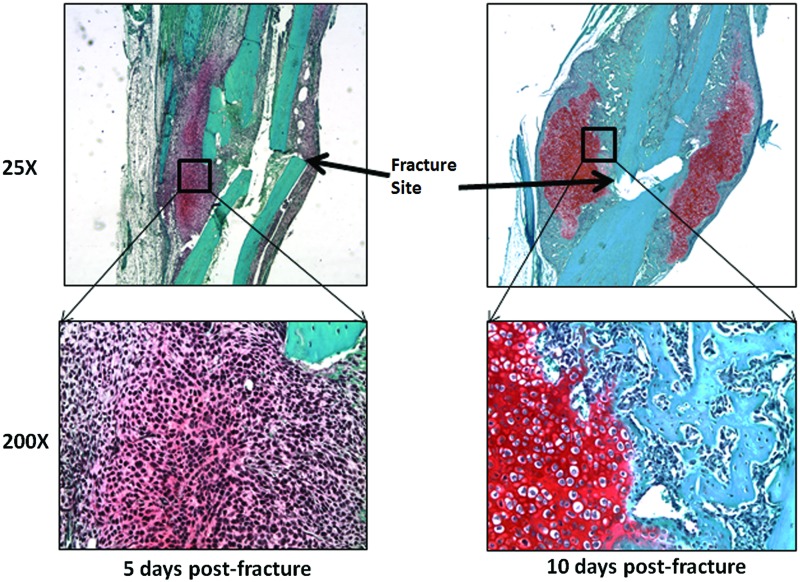
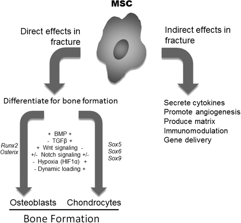
Significance: Mesenchymal stem cells (MSCs) play a key role in fracture repair by differentiating to become bone-forming osteoblasts and cartilage-forming chondrocytes. Cartilage then serves as a template for additional bone formation through the process of endochondral ossification.
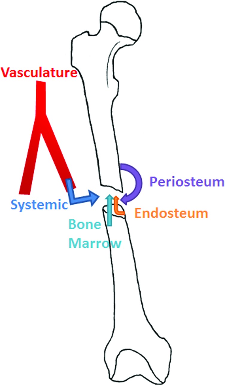
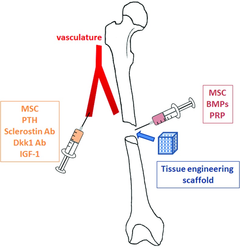
Recent advances: Endogenous MSCs that contribute to healing are primarily derived from the periosteum, endosteum, and marrow cavity, but also may be contributed from the overlying muscle or through systemic circulation, depending on the type of injury. A variety of growth factor signaling pathways, including BMP, Wnt, and Notch signaling, influence MSC proliferation and differentiation. These MSCs can be therapeutically manipulated to promote differentiation. Furthermore, MSCs can be harvested, cultivated, and delivered to promote bone healing.
Critical issues: Pharmacologically manipulating the number and differentiation capacity of endogenous MSCs is one potential therapeutic approach to improve healing; however, ideal agents to influence signaling pathways need to be developed and additional therapeutics that activate endogenous MSCs are needed. Whether isolated and purified, MSCs participate directly in the healing process or serve a bystander effect and indirectly influence healing is not well defined.
Future directions: Studies must focus on better understanding the regulation of endogenous MSCs durings fracture healing. This will reveal novel molecules and pathways to therapeutically target. Similarly, while animal models have demonstrated efficacy in the delivery of MSCs to promote healing, more research is needed to understand ideal donor cells, cultivation methods, and delivery before stem cell therapy approaches can be utilized to repair bone.
Figures
References
-
- Cohnheim J. Über entzündung und eiterung. Path Anat Physiol Klin Med. 1867;40:1.
-
- Goujon E. Recherches experimentales sue les proprieties. J L Anat. 1869;6:399.
-
- Friedenstein AJ. Chailakhjan RK. Lalykina KS. The development of fibroblast colonies in monolayer cultures of guinea-pig bone marrow and spleen cells. Cell Tissue Kinet. 1970;3:393. - PubMed
-
- Caplan AI. Mesenchymal stem cells. J Orthop Res. 1991;9:641. - PubMed
Publication types
LinkOut - more resources
Full Text Sources
Other Literature Sources
