Review
. 2013 Sep;2(7):379-388.
doi: 10.1089/wound.2012.0383.

Neutrophils and Wound Repair: Positive Actions and Negative Reactions

Affiliations
Review

Neutrophils and Wound Repair: Positive Actions and Negative Reactions

Traci A Wilgus et al. Adv Wound Care (New Rochelle). 2013 Sep.

Abstract

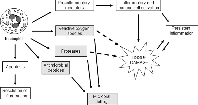
Significance: Neutrophils are one of the most abundant cells of the immune system and they are extremely active during the repair of cutaneous wounds. In general, the antimicrobial activity of neutrophils is effective and allows these cells to carry out their primary function of preventing wounds from becoming infected.

Recent advances: It is now known that in addition to sterilizing the wound, the weapons used by neutrophils to kill potential pathogens can also cause significant tissue damage to the host. This additional damage can lead to delayed healing and excessive scar formation.

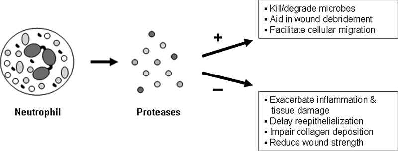
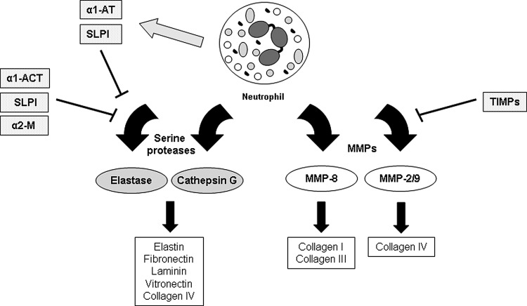
Critical issues: Much of the host damage caused by neutrophils results from the activity of proteases secreted by these cells. The clinical significance of this problem is highlighted by numerous studies showing that high levels of neutrophil-derived proteases are associated with chronic, non-healing wounds.

Future directions: Studies are currently being performed to evaluate new ways of counteracting protease activity in chronic wounds. Additional studies will have to be carried out to determine whether neutralizing neutrophil proteases can improve the healing of chronic wounds without sacrificing the ability of neutrophils to eliminate pathogens and risking infection.


Figures

None
Traci A. Wilgus, PhD
Figure 1.
Figure 1.
Overview of neutrophil activities during cutaneous repair. Circulating neutrophils are recruited to the wound site quickly after injury, where they carry out a variety of functions. They produce an array of proinflammatory mediators that recruit and activate other inflammatory cells, enhancing inflammation. In normal wounds, recruited neutrophils will eventually undergo apoptosis and be engulfed by macrophages, initiating a resolution program that terminates the inflammatory response. However, in non-healing wounds, inflammatory cells often continue to be recruited and activated, leading to persistent inflammation. Activated neutrophils help prevent wound infection by generating reactive oxygen species and producing proteases and antimicrobial peptides. These substances kill and degrade potentially pathogenic microbes, but reactive oxygen species and proteases, in particular, can cause tissue damage when released extracellularly. Excessive proteases can be especially harmful, causing unwanted degradation of the extracellular matrix and additional tissue damage. This can lead to a harmful cycle, whereby the damage caused by neutrophil-derived proteases causes even more inflammation. This leads to more tissue damage, eventually stalling the wound-healing process and preventing complete closure of the wound.
Figure 2.
Figure 2.
Neutrophil recruitment in the tissue adjacent to an acute murine skin wound. Ly-6G immunostaining is commonly used to detect tissue neutrophils (brown staining). Neutrophils are not frequently observed in normal skin of 6-week-old FVB mice (left). However, damage to the skin sets in motion a series of events causing neutrophils to travel from the bloodstream to the site of injury. This is a rapid process, and a large number of neutrophils can be seen at the wound margin, or the tissue immediately adjacent to the wound bed (WB), after injury (right). The image shows skin adjacent to a full-thickness incisional FVB mouse wound at 48 h post-injury. Open arrows are used to indicate areas heavily populated with neutrophils (brown cells). E, epidermis; D, dermis; M, muscle; WB, wound bed. To see this illustration in color, the reader is referred to the web version of this article at www.liebertpub.com/wound
Figure 3.
Figure 3.
Neutrophil maturation and granule formation. Mature neutrophils have a variety of granule types that contain distinct sets of mediators involved in neutrophil function. Neutrophil precursors originate in the bone marrow and undergo differentiation in the blood. The formation of different granule types coincides with specific stages of neutrophil development. Azurophilic granules begin to form at the promyelocyte stage, specific granule formation spans the myelocyte and metamyelocyte stages, and gelatinase granules are produced in metamyelocytes and band cells. Secretory vesicles form last through endocytosis. Because gene expression patterns change during the course of neutrophil development, each granule type contains a distinct set of proteins. During an inflammatory response, mature neutrophils leave the bloodstream by undergoing extravasation. After traversing the endothelial cell lining of blood vessels, neutrophils enter the tissue and migrate to the site of inflammation or injury.
Figure 4.
Figure 4.
Beneficial and detrimental effects of neutrophil-derived proteases on wound healing. Activated neutrophils produce a number of proteases that can have positive (+) or negative (−) effects on the cutaneous repair process. Neutrophil-derived proteases help kill and degrade microbes and break down components of the extracellular matrix, which can debride the wound and facilitate cellular migration when present at appropriate levels. Conversely, excessive levels of proteases in the tissue resulting from overly active neutrophils or prolonged neutrophil recruitment can be detrimental by causing additional tissue damage and further inflammation. Consequently, defective collagen deposition, reduced wound strength, and delayed re-epithelialization can occur.
Figure 5.
Figure 5.
Neutrophil proteases involved in wound healing and their inhibitors. Neutrophils produce two main classes of proteases relevant to tissue repair. Serine proteases like elastase and cathepsin G target a variety of extracellular matrix proteins, including elastin, fibronectin, laminin, vitronectin, and collagen IV. The action of serine proteases is balanced by several protease inhibitors produced by neutrophils (α1-AT, SLPI) and surrounding skin cells (α1-ACT, SLPI, α2-M). Neutrophils also produce several types of MMPs. MMP-8, which cleaves fibrillar collagen, and MMP-2/MMP-9, which cleave collagen IV (among other substrates), are involved in wound repair. The activity of MMPs is inhibited by a class of molecules called TIMPs produced by a variety of cells in the skin. If the activity of proteases and their inhibitors is not tightly regulated, the protease activity can become extreme and impair the healing process.

References

    1. Theilgaard-Monch K. Knudsen S. Follin P. Borregaard N. The transcriptional activation program of human neutrophils in skin lesions supports their important role in wound healing. J Immunol. 2004;172:7684. - PubMed
    1. Nathan C. Neutrophils and immunity: challenges and opportunities. Nat Rev Immunol. 2006;6:173. - PubMed
    1. Serhan CN. Chiang N. Van Dyke TE. Resolving inflammation: dual anti-inflammatory and pro-resolution lipid mediators. Nat Rev Immunol. 2008;8:349. - PMC - PubMed
    1. Widgerow AD. Cellular resolution of inflammation—catabasis. Wound Repair Regen. 2012;20:2. - PubMed
    1. Gutierrez-Fernandez A. Inada M. Balbin M. Fueyo A. Pitiot AS. Astudillo A. Hirose K. Hirata M. Shapiro SD. Noel A. Werb Z. Krane SM. Lopez-Otin C. Puente XS. Increased inflammation delays wound healing in mice deficient in collagenase-2 (MMP-8) FASEB J. 2007;21:2580. - PMC - PubMed