. 2014 Jan 30;2(2):127-34.
doi: 10.1016/j.stemcr.2013.12.012. eCollection 2014 Feb 11.

Wnt secretion from epithelial cells and subepithelial myofibroblasts is not required in the mouse intestinal stem cell niche in vivo

Affiliations

Wnt secretion from epithelial cells and subepithelial myofibroblasts is not required in the mouse intestinal stem cell niche in vivo

Adrianna K San Roman et al. Stem Cell Reports. .

Abstract

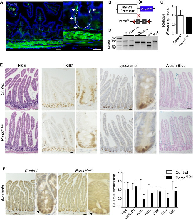
Wnt signaling is a crucial aspect of the intestinal stem cell niche required for crypt cell proliferation and differentiation. Paneth cells or subepithelial myofibroblasts are leading candidate sources of the required Wnt ligands, but this has not been tested in vivo. To abolish Wnt-ligand secretion, we used Porcupine (Porcn) conditional-null mice crossed to strains expressing inducible Cre recombinase in the epithelium, including Paneth cells (Villin-Cre (ERT2) ); in smooth muscle, including subepithelial myofibroblasts (Myh11-Cre (ERT2) ); and simultaneously in both compartments. Elimination of Wnt secretion from any of these compartments did not disrupt tissue morphology, cell proliferation, differentiation, or Wnt pathway activity. Thus, Wnt-ligand secretion from these cell populations is dispensable for intestinal homeostasis, revealing that a minor cell type or significant and unexpected redundancy is responsible for physiologic Wnt signaling in vivo.


Figures

None
Graphical abstract
Figure 1
Figure 1
Porcn Deletion in Epithelial Cells Spares Intestinal Epithelial Morphology and Function (A) Strategy for Porcn deletion by TAM-inducible Cre recombinase driven by epithelium-specific Villin promoter. ER, estrogen receptor. (B) Genomic PCR of epithelium isolated from four PorcnE-Del intestines indicates complete recombination of the floxed allele (485 bp band) compared to two Porcn+/Y control mice (685 bp band from the wild-type allele). For reference, DNA is also shown from single PorcnDel/X (Δ/+; 485 bp and 685 bp products) and PorcnFl/Y;Cre− (762 bp band from the unrecombined floxed allele) mice. WT, wild-type. (C) qRT-PCR analysis of Porcn mRNA in isolated PorcnE-Del intestinal crypts (N = 4) reveals reduction by orders of magnitude compared to controls (N = 2). (D) Histology and immunohistochemistry (IHC) of PorcnE-Del (N = 5) and control (Porcn+/Y;Villin-CreERT2; N = 3) mice reveals no abnormalities. Left to right: hematoxylin and eosin (H&E) staining, Ki67 IHC, lysozyme IHC, and Alcian blue staining. High-magnification images are shown to the right of low-magnification views. (E) Left, β-catenin IHC in control and PorcnE-Del mice shows nuclear-staining crypt-base cells (arrows). Right, qRT-PCR analysis of Wnt target mRNAs in isolated control (N = 2) and PorcnE-Del (N = 4) crypt epithelium demonstrates unperturbed Wnt-pathway activity. Bars represent mean ± SEM of biological replicates; all scale bars, 50 μm.
Figure 2
Figure 2
Porcn Loss in Intestinal Smooth Muscle Cells and SEMFs Does Not Adversely Affect Epithelial Morphology and Function (A) Myh11-CreERT2;Rosa26R mice activate Cre recombinase in all muscle layers, including subepithelial myofibroblasts. The dotted line in the high-magnification image (right) outlines a crypt, and arrows point to the thin SEMF layer of YFP+ cells enveloping the crypt. The scale bars represent 30 μm. See also Figure S2. (B) Strategy to induce muscle-cell-specific recombination in PorcnFl/Y;Myh11-CreERT2 mice. (C) qRT-PCR analysis of isolated crypt epithelium shows no significant difference in Porcn mRNA expression in PorcnM-Del (N = 4) mice compared to control (Porcn+/Y;Myh11-CreERT2, N = 4) mice, p = 0.35. (D) PCR from whole-intestine genomic DNA, revealing the expected proportion of recombined Porcn in smooth muscle, with residual unrecombined Porcn DNA contributed by epithelial and other cell types that lack Myh11-CreERT2 expression. (E) Histology and immunostains on PorcnM-Del (N = 7) and control (Porcn+/Y;Myh11-CreERT2, N = 6) intestines reveal no abnormalities. (F) Left, β-catenin IHC in control and PorcnM-Del mice shows nuclear staining in crypt-base cells (arrows). Right, qRT-PCR reveals intact Wnt target expression in crypts isolated from PorcnM-Del intestines, compared to controls (N = 4; statistics in Table S1). Bars represent mean ± SEM of biological replicates; scale bars (E and F), 50 μm.
Figure 3
Figure 3
Consequences of Simultaneous Loss of Porcn from Epithelial and Muscle Cells (A) DNA from whole intestines in TAM-treated PorcnFl/Y;Villin-CreERT2;Myh11-CreERT2 mice shows significant recombination at the floxed Porcn allele, with a minimal contribution of unrecombined DNA from nonepithelial, nonmuscle cells. (B) Left, qRT-PCR analysis of RNA from crypts of PorcnEM-Del mutants (N = 3) compared to controls (Porcn+/Y;Villin-CreERT2;Myh11-CreERT2; N = 3) shows total Porcn deficiency, p = 0.0013. Right, qRT-PCR of unfractionated PorcnM-Del (N = 2) intestines shows lower Porcn expression than controls (Porcn+/Y;Villin-CreERT2;Myh11-CreERT2 and Porcn+/Y;Myh11-CreERT2; N = 4), which is further reduced in PorcnEM-Del (N = 2) mice. (C) Equivalent BrdU uptake in control and PorcnEM-Del intestinal crypts. (D) Histology and immunostains from PorcnEM-Del and control intestines reveal intact morphology, cell proliferation, and differentiation (N = 3). (E) Left, β-catenin IHC in control and PorcnEM-Del mice shows nuclear staining in crypt-base cells (arrows). Right, qRT-PCR analysis of control (N = 3) and PorcnEM-Del (N = 3) intestines indicates a statistically insignificant (Table S1) reduction in levels of Wnt target transcripts. Bars in graphs represent mean ± SEM of biological replicates; all scale bars, 50 μm.

References

    1. Barker N., van Es J.H., Kuipers J., Kujala P., van den Born M., Cozijnsen M., Haegebarth A., Korving J., Begthel H., Peters P.J., Clevers H. Identification of stem cells in small intestine and colon by marker gene Lgr5. Nature. 2007;449:1003–1007. - PubMed
    1. Barker N., Ridgway R.A., van Es J.H., van de Wetering M., Begthel H., van den Born M., Danenberg E., Clarke A.R., Sansom O.J., Clevers H. Crypt stem cells as the cells-of-origin of intestinal cancer. Nature. 2009;457:608–611. - PubMed
    1. Barrott J.J., Cash G.M., Smith A.P., Barrow J.R., Murtaugh L.C. Deletion of mouse Porcn blocks Wnt ligand secretion and reveals an ectodermal etiology of human focal dermal hypoplasia/Goltz syndrome. Proc. Natl. Acad. Sci. USA. 2011;108:12752–12757. - PMC - PubMed
    1. Bastide P., Darido C., Pannequin J., Kist R., Robine S., Marty-Double C., Bibeau F., Scherer G., Joubert D., Hollande F. Sox9 regulates cell proliferation and is required for Paneth cell differentiation in the intestinal epithelium. J. Cell Biol. 2007;178:635–648. - PMC - PubMed
    1. Biechele S., Cox B.J., Rossant J. Porcupine homolog is required for canonical Wnt signaling and gastrulation in mouse embryos. Dev. Biol. 2011;355:275–285. - PubMed

Publication types

MeSH terms