. 2014 May;99(5):E813-20.
doi: 10.1210/jc.2013-3186. Epub 2014 Feb 14.

Signaling through FSH receptors on human umbilical vein endothelial cells promotes angiogenesis

Affiliations

Signaling through FSH receptors on human umbilical vein endothelial cells promotes angiogenesis

Julie A Stilley et al. J Clin Endocrinol Metab. 2014 May.

Abstract

Context: The FSH receptor (FSHR) is traditionally thought to play a role in female reproductive physiology solely within the context of ovarian FSHR. However, FSHR is also expressed in endothelial cells of the placental vasculature and human umbilical cord vessels, suggesting additional facets of female reproduction regulated by extragonadal FSHR.

Objective: We sought to determine the functional role of FSHR on human umbilical cord endothelial cells (HUVECs), hypothesizing that activation of the FSHR would stimulate angiogenesis.

Design: The ability of FSH to stimulate several angiogenic processes in HUVECs was determined.

Setting: This was a laboratory-based study using commercially prepared HUVECs.

Results: Tube formation, wound healing, cell migration, cell proliferation, nitric oxide production, and cell survival were stimulated in response to FSH. Quantitative comparisons between HUVECs incubated with maximally stimulatory concentrations of FSH vs vascular endothelial growth factor (VEGF), a well-characterized angiogenic factor, revealed that FSH is as efficacious as VEGF in promoting angiogenic processes. FSH did not provoke increased secretion of VEGF by HUVECs, suggesting the direct stimulation of angiogenic processes by FSH in endothelial cells. In contrast to gonadal cells, the FSHR on HUVECs did not mediate an FSH-stimulated increase in cAMP. However, increased phosphorylation of AKT in response to FSH was observed, suggesting that FSH stimulation of HUVEC FSHR stimulates the PI3K/AKT signaling pathway.

Conclusions: Our studies reveal a novel role for FSHR in female reproductive physiology. Its ability to promote angiogenesis in placental endothelial cells suggests that the FSHR may have an influential role in pregnancy.


Figures

Figure 1.
Figure 1.
FSHR is detected immunohistochemically on endothelial cells of human umbilical cord vein and on HUVECs. The vein of human umbilical cord from term pregnancy was stained with anti-FSHR323 (A) or with isotype- and concentration-matched nonimmune IgG (B). A and B, Magnification ×200. Labeled are the tunica interna (TI), tunica media (TM), and Whartson's Jelly (WJ). Immunofluorescence of HUVECs stained with anti-FSHR323 and counterstained with DAPI (blue) (C) or with isotype- and concentration-matched nonimmune IgG and counterstained with DAPI (blue) (D). The immunohistochemical staining of FSHR in human umbilical cord is representative of that observed in at least six different cords. The immunofluorescence imaging of FSHR in HUVECs is representative of three independent experiments.
Figure 2.
Figure 2.
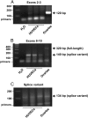
FSHR mRNA in HUVECs. cDNA was extended using end-point PCR. Traditional PCR amplifying exons 2 though 3 (A), gene-specific PCR amplifying exons 8 through 10 (B), and gene-specific PCR amplifying an FSHR mRNA splice variant lacking exon 9 (C) were performed as described in the Supplemental Data. Shown are the results using three different cultures of pooled HUVECs and from pooled human ovaries. Data shown are representative of three independent experiments.
Figure 3.
Figure 3.
FSH stimulates tube formation of HUVECs. Serum-starved HUVECs plated on a reduced growth factor basement membrane matrix were incubated with no additions (A), FSH (600 ng/mL final concentration) (B), or LSGS (C). Data are representative of three independent experiments. Dose response curves quantifying tube length as a function of final concentrations of FSH (D) or VEGF (E) are shown. Data shown in D and E are each the mean ± SEM of triplicate determinations of a single experiment and each are representative of two individual experiments.
Figure 4.
Figure 4.
FSH stimulates several angiogenic processes in HUVECs with a similar efficacy as VEGF. Within a given experiment in each assay, serum-starved HUVECs were incubated with vehicle (no additions [NA]), FSH at 600 ng/mL final concentration, VEGF at 50 ng/mL final concentration, or LSGS, and the following assays were performed and quantified: tube formation (A), wound healing (B), cell migration (C), and cell proliferation as determined by Click-iT EdU (D). Data shown are the mean ± SEM of five, three, three, and four independent experiments in A, B, C, and D, respectively. Asterisks denote differences with P < .5.
Figure 5.
Figure 5.
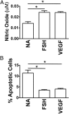
FSH stimulates nitric oxide production and promotes cell survival in HUVECs. Serum-starved HUVECs were incubated with vehicle (no additions [NA]), FSH at 600 ng/mL final concentration, or VEGF at 50 ng/mL final concentration. Nitric oxide production (A) and antiapoptotic effects (B) were quantified. Data shown are the mean ± SEM of four and three independent experiments in A and B, respectively. Asterisks denote differences with P < .5.
Figure 6.
Figure 6.
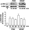
FSH stimulates Akt phosphorylation in HUVECs. Serum-starved HUVECs were incubated for the indicated times with FSH at 600 ng/mL final concentration or VEGF at 50 ng/mL final concentration. Total Akt (t-Akt) and phosphorylated Akt (p-Akt) were determined by Western blotting. A, Western blots from one representative experiment. B, The ratio of p-Akt/t-Akt was determined and plotted as the fold-increase relative to time zero. Data shown are the mean ± SEM of three independent experiments.

Similar articles

Cited by

References

    1. Khankin EV, Royle C, Karumanchi SA. Placental vasculature in health and disease. Semin Thromb Hemost. 2010;36:309–320 - PubMed
    1. Young BC, Levine RJ, Karumanchi SA. Pathogenesis of preeclampsia. Annu Rev Pathol. 2010;5:173–192 - PubMed
    1. Radu A, Pichon C, Camparo P, et al. Expression of follicle-stimulating hormone receptor in tumor blood vessels. N Engl J Med. 2010;363:1621–1630 - PubMed
    1. Zhu LL, Blair H, Cao J, et al. Blocking antibody to the β-subunit of FSH prevents bone loss by inhibiting bone resorption and stimulating bone synthesis. Proc Natl Acad Sci USA. 2012;109:14574–14579 - PMC - PubMed
    1. Zhang M, Tao YX, Ryan GL, Feng X, Fanelli F, Segaloff DL. Intrinsic differences in the response of the human lutropin receptor versus the human follitropin receptor to activating mutations. J Biol Chem. 2007;282:25527–25539 - PubMed

Publication types

MeSH terms