

Review
doi: 10.1016/j.ceb.2013.10.006.
Epub 2013 Nov 16.
Morphology and function of membrane-bound organelles
Affiliations
- PMID: 24529249
- PMCID: PMC3927147
- DOI: 10.1016/j.ceb.2013.10.006
Item in Clipboard
Review
Morphology and function of membrane-bound organelles
Curr Opin Cell Biol.
2014 Feb.
Abstract
The cell interior is a busy and crowded place. A large fraction of the cell volume is taken up by organelles that come in a variety of shapes and sizes. These organelles are surrounded by membrane that not only acts as a diffusion barrier, but also provides each organelle with its unique morphology that contributes to its function, often in ways that are poorly understood. Here we discuss recent discoveries on the relationship between organelle structure and function.
Copyright © 2013 Elsevier Ltd. All rights reserved.
Figures
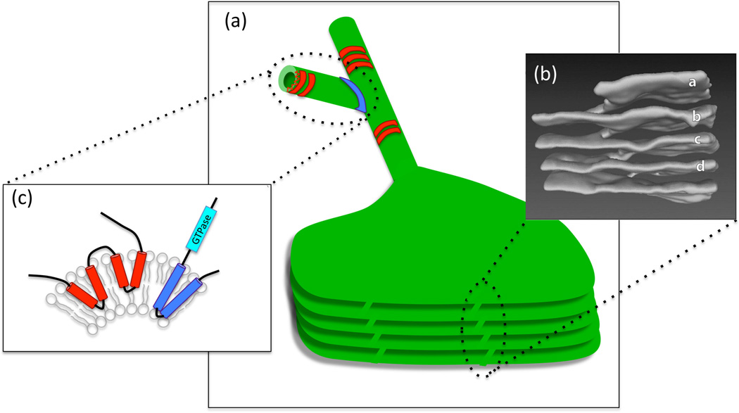
Diverse membrane structures in the ER. (a) The ER is an interconnected network of composed of branched tubules and sheets, some of which can form stacks, as shown in the illustration. ER tubules are stabilized by the oligomerization of proteins such as reticulons and DP1/Yop1/REEps (in red), while 3-way junctions are mediated by proteins such as atlastins (in blue). (b) The structure of reticulons and atlastin. Membrane curvature is induced by the insertion of protein "wedges" (two in the case of reticulons and one in the case of atlastin) that traverse only one lipid bilayer, forcing the membrane to curve. Atlastin has an in addition GTPase activity that is necessary for fusing membranes and generating 3-way junctions. (c) Helicoidal membrane structure in stacked ER sheets. A 3D reconstruction of a region of stacked ER sheets from an acinar cell of a mouse salivary gland. The letters (a through d) mark the ER sheets that are connected through a helicoidal structure (to the left). From Terasaki et al. Cell 154, 285–296, 2013.
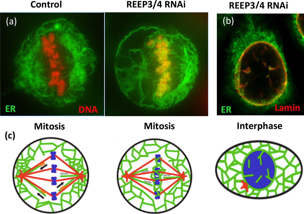
Depletion of REEP3/4 causes accumulation of ER on mitotic chromosomes and leads to intranuclear membranes and lamina. A. HeLa cells expressing RFP-histone (red) to label the DNA and GFP-Sec61 (green) to mark the ER were subjected to control or REEP3/4 RNAi and imaged during mitosis. Note the colocalization of ER and mitotic chromosomes in the absence of REEP3/4. (b) An interphase REEP3/4 RNAi-treated HeLa cell expressing GFP-Sec61 (green) was fixed and stained for nuclear lamin B1 (red). Both NE markers are aberrantly localized to structures inside the nucleus. (c) Schematic of phenotypes with microtubules in red, DNA in blue and ER in green. Adapted from Schlaitz et al. Dev. Cell 26, 316–323, 2013. Images courtesy of Anne-Lore Schlaitz and Rebecca Heald.
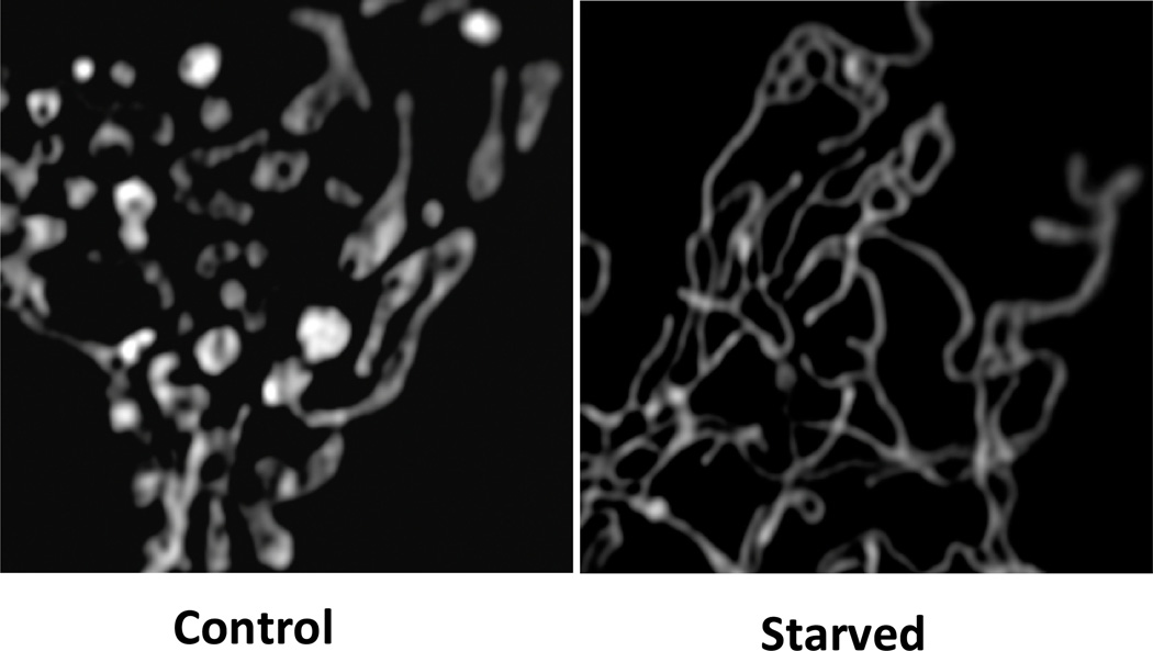
Starvation leads to unopposed mitochondrial fusion. Mouse embryo fibroblasts transfected with mitoRFP were incubated in full nutrient medium (Control), or starvation medium (Starved) for 6 hours. Starved cells show a continuous network of mitochondria. Cells were fixed and images acquired by Structured Illumination Microscopy. Images courtesy of Angelika Rambold and Jennifer Lippencott-Schwartz.
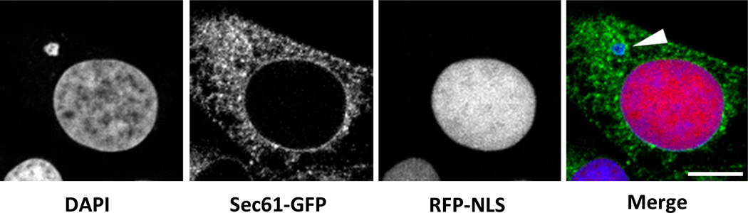
Micronuclei are unstable in somatic cells. The images show a U2OS cell containing an intact nucleus and a disrupted micronucleus (arrowhead). The micronucleus fails to accumulate the fluorescent nuclear protein RFP-NLS, and has been invaded by ER as indicated by the presence of Sec61-GFP. Scale bar = 10 microns. Images courtesy of Emily Hatch and Martin Hetzer.
References
-
- Bereiter-Hahn J, Jendrach M. Mitochondrial dynamics. Int Rev Cell Mol Biol. 2010;284:1–65. - PubMed
Publication types
MeSH terms
Grants and funding
LinkOut - more resources
Full Text Sources
Other Literature Sources
