. 2014 Feb 13;156(4):705-16.
doi: 10.1016/j.cell.2014.01.019.

A promiscuous lipid-binding protein diversifies the subcellular sites of toll-like receptor signal transduction

Affiliations

A promiscuous lipid-binding protein diversifies the subcellular sites of toll-like receptor signal transduction

Kevin S Bonham et al. Cell. .

Abstract

The Toll-like receptors (TLRs) of the innate immune system are unusual in that individual family members are located on different organelles, yet most activate a common signaling pathway important for host defense. It remains unclear how this common signaling pathway can be activated from multiple subcellular locations. Here, we report that, in response to natural activators of innate immunity, the sorting adaptor TIRAP regulates TLR signaling from the plasma membrane and endosomes. TLR signaling from both locations triggers the TIRAP-dependent assembly of the myddosome, a protein complex that controls proinflammatory cytokine expression. The actions of TIRAP depend on the promiscuity of its phosphoinositide-binding domain. Different lipid targets of this domain direct TIRAP to different organelles, allowing it to survey multiple compartments for the presence of activated TLRs. These data establish how promiscuity, rather than specificity, can be a beneficial means of diversifying the subcellular sites of innate immune signal transduction.


Figures

Figure 1
Figure 1. TIRAP is required for endosomal TLR signaling in response to natural ligands
(A, B) Primary WT or TIRAP KO BMDM were treated with 100ng/mL LPS or 1μM CpG DNA (A) or infected with HSV (B) for 3 hours. Total mRNA was extracted and analyzed for expression of IL-1β and IL-6. Note that TIRAP KO BMDM are defective in responding to natural ligands. (C) Microarray gene expression profiles from sorted cDCs and pDCs were analyzed for transcripts associated with TLR-signaling pathways. Values on the x-axis represent the fold difference in gene expression between mDCs and pDCs. Grey area depicts equal gene expression in cDCs and pDCs. Note that TIRAP is expressed in pDCs comparably to cDCs, despite the absence of cell-surface TLRs. (D) pDCs from WT or TIRAP KO mice were infected with HSV-2 (186 syn+) at MOI=1 or influenza (A/PR/8) at MOI=10. Supernatants were collected after 24 hours and analyzed by ELISA. Error bars represent SD. See also Figure S1.
Figure 2
Figure 2. Immortalized BMDM responses to CpG DNA mimic primary cell responses to natural ligands
(A) WT, TIRAP KO or TIRAP-expressing TIRAP KO iBMDM were infected with indicated HSV strains (MOI=1) for 3 hours and analyzed by qPCR. (B) Primary or iBMDM were incubated with fluorescent beads at 37°C for the indicated times and phagocytosis was analyzed by flow cytometry. Mean fluorescence intensity was plotted relative to cells incubated with beads on ice for 45 min. (C) WT or TIRAP KO primary and iBMDM were stimulated with 1μM CpG DNA. After 24 hours, supernatants were collected and analyzed for IL-6 by ELISA. N.D. indicates that no signal was detected. (D) WT, TIRAP KO or TIRAP-expressing TIRAP KO iBMDM were treated with LPS or CpG DNA for 3 hours and analyzed by qPCR. Error bars represent SD.
Figure 3
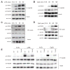
Figure 3. TIRAP is a critical constituent of the myddosome
(A–B) WT iBMDM were stimulated with LPS (A) or CpG DNA (B) for the indicated times. MyD88 was immunoprecipitated from lysates and analyzed by western blot. (C–D) TIRAP-transgenic iBMDM were stimulated with LPS (C) or CpG DNA (D) and biotin-TIRAP was precipitated from cleared lysates with avidin-coated agarose before western analysis. (E) WT, TIRAP KO or TIRAP-expressing TIRAP KO iBMDM were treated with LPS for 1 hour or CpG DNA for two hours and analyzed as in (A). N.S. indicates cells that were not stimulated. See also Figure S2.
Figure 4
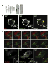
Figure 4. Promiscuous lipid binding diversifies the localization of TIRAP
(A) GST-tagged TIRAP or TIRAP 4X proteins were incubated with PIP strips containing various lipids (shown in left panel) and assessed for protein binding by far western analysis. (B) TIRAP KO primary BMDM expressing TIRAP loc-GFP and PLC-cherry were analyzed by confocal microscopy. All images are representative of at least three independent experiments where over 200 cells were examined per condition and >95% of the cells displayed similar staining. Selective localization of TIRAP to intracellular vesicles compared to the PLCδ1 PH domain demonstrates that TIRAP binds to multiple lipids. (C) TIRAP KO primary BMDM expressing TIRAP-loc GFP were analyzed by confocal microscopy. One image was captured every 20 seconds for 20 min. (C) Shows representative frames from one such capture (See Movie S1 for full-length movie).
Figure 5
Figure 5. Promiscuous lipid binding by TIRAP diversifies the sites of TLR signaling
(A) Confocal microscopic analysis of TIRAP KO iBMDM stably transduced with GFP-tagged TIRAP alleles that contain different lipid binding domains (depicted in the top panel). The micrographs demonstrate selective localization of TIRAP dependent on its lipid binding domain. All images are representative of at least three independent experiments where over 200 cells were examined per condition and >95% of the cells displayed similar staining. (B) Cells from (A) were stimulated with LPS or CpG DNA and analyzed by qPCR. Note that selective binding of TIRAP to distinct lipids permits signaling from distinct compartments. (C) Cells from (A) were stimulated with LPS or CpG DNA for one hour and myddosome formation was assessed. (D) Model depicting how the promiscuous lipid-binding domain of TIRAP promotes signal transduction from the plasma membrane and endosomes. TLRs found at the cell surface signal from a PI(4,5)P2-rich subdomain, and TIRAP is recruited to that location via interactions with PI(4,5)P2. TLRs found on endosomes signal from a domain rich in 3′ phosphoinositides (depicted is PI(3)P). These lipids direct TIRAP to endosomes to promote TLR signaling. Error bars represent SD.

Comment in

References

    1. Akira S, Uematsu S, Takeuchi O. Pathogen recognition and innate immunity. Cell. 2006;124:783–801. - PubMed
    1. Aksoy E, Taboubi S, Torres D, Delbauve S, Hachani A, Whitehead MA, Pearce WP, Berenjeno-Martin I, Nock G, Filloux A, et al. The p110delta isoform of the kinase PI(3)K controls the subcellular compartmentalization of TLR4 signaling and protects from endotoxic shock. Nat Immunol. 2012;13:1045–1054. - PMC - PubMed
    1. Baeuerle PA, Baltimore D. Activation of DNA-binding activity in an apparently cytoplasmic precursor of the NF-kappa B transcription factor. Cell. 1988;53:211–217. - PubMed
    1. Barbalat R, Ewald SE, Mouchess ML, Barton GM. Nucleic acid recognition by the innate immune system. Annu Rev Immunol. 2011;29:185–214. - PubMed
    1. Barbalat R, Lau L, Locksley RM, Barton GM. Toll-like receptor 2 on inflammatory monocytes induces type I interferon in response to viral but not bacterial ligands. Nat Immunol. 2009;10:1200–1207. - PMC - PubMed

Publication types

MeSH terms

Associated data