. 2014 Feb 13;156(4):800-11.
doi: 10.1016/j.cell.2014.01.021.

A bicistronic MAVS transcript highlights a class of truncated variants in antiviral immunity

Affiliations

A bicistronic MAVS transcript highlights a class of truncated variants in antiviral immunity

Sky W Brubaker et al. Cell. .

Abstract

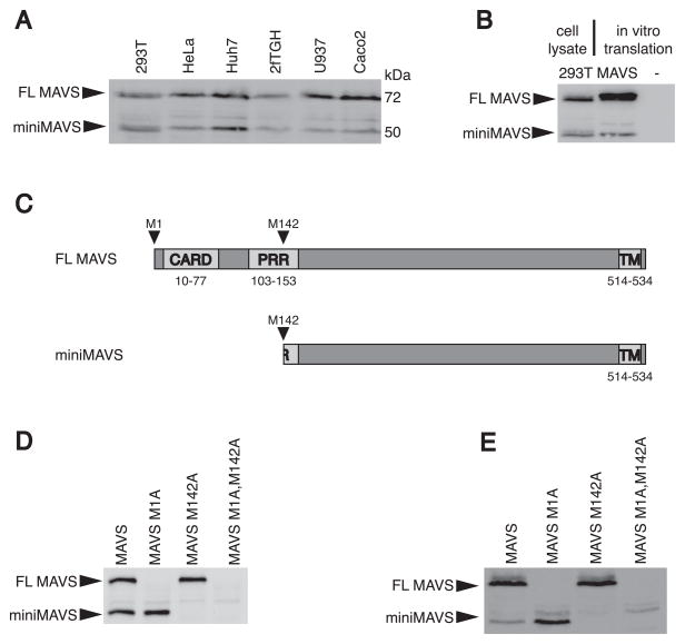
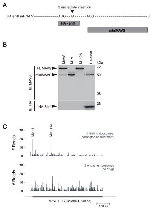
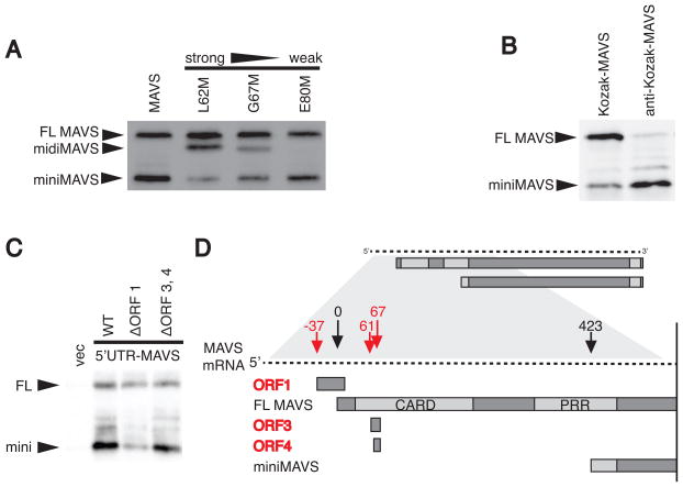
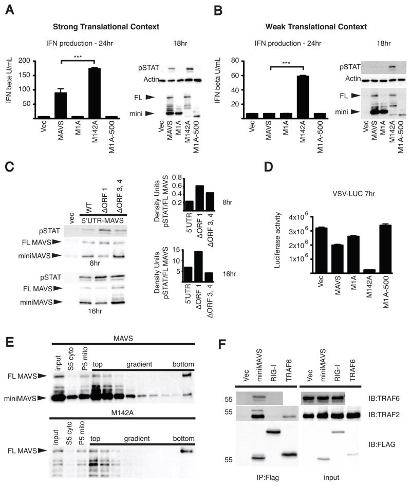
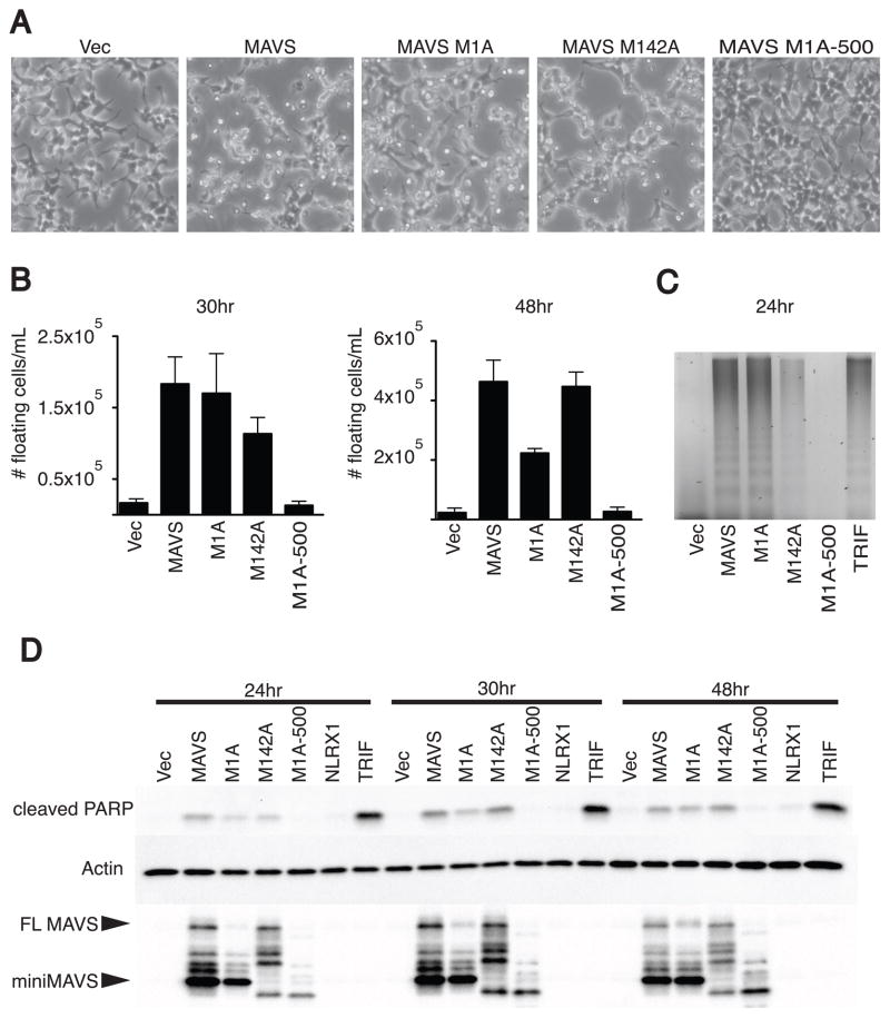
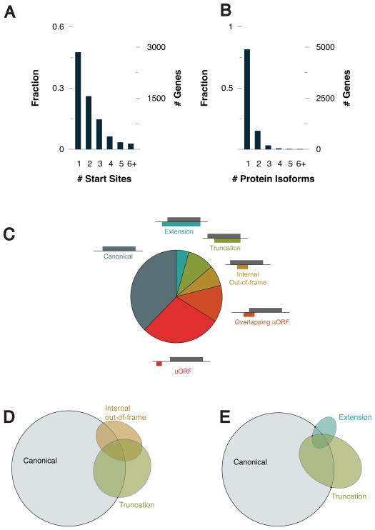
Bacterial and viral mRNAs are often polycistronic. Akin to alternative splicing, alternative translation of polycistronic messages is a mechanism to generate protein diversity and regulate gene function. Although a few examples exist, the use of polycistronic messages in mammalian cells is not widely appreciated. Here we report an example of alternative translation as a means of regulating innate immune signaling. MAVS, a regulator of antiviral innate immunity, is expressed from a bicistronic mRNA encoding a second protein, miniMAVS. This truncated variant interferes with interferon production induced by full-length MAVS, whereas both proteins positively regulate cell death. To identify other polycistronic messages, we carried out genome-wide ribosomal profiling and identified a class of antiviral truncated variants. This study therefore reveals the existence of a functionally important bicistronic antiviral mRNA and suggests a widespread role for polycistronic mRNAs in the innate immune system.


Figures

Figure 1
Figure 1. miniMAVS is expressed from a second translational start site
(A) Lysates from several different human cell lines were separated by SDS-PAGE and endogenous MAVS expression was detected with a MAVS antibody. (B) in vitro transcription and translation of the MAVS CDS was compared with 293T cell lysates with an anti-MAVS antibody. (C) Schematic of MAVS with predicted translation products FL MAVS and miniMAVS from the start sites corresponding to Met 1 and Met 142. (D–E) Point mutations of translational start sites at Met 1 and Met 142 were made in the MAVS CDS and expressed in vitro (D) and in vivo (E) from MAVS deficient MEFs. The translation products were detected by immunoblot with a MAVS antibody. See also Figure S1.
Figure 2
Figure 2. MAVS is bicistronic and in vivo ribosome initiation is detected at the FL MAVS and miniMAVS start sites
(A) Schematic of the HA-shift expression vector containing a frame shift mutation and the predicted translation products “HA-shift” and miniMAVS. (B) Lysates from stable MEF lines expressing the MAVS and HA-shift constructs were separated by SDS-PAGE and protein expression was determined with MAVS and HA antibodies. (C) Pattern of ribosome initiation (harringtonine treatment) and elongation on endogenous MAVS mRNA in 293T cells.
Figure 3
Figure 3. Cis-regulatory elements of the MAVS transcript control miniMAVS expression
(A) Translational start sites of varying strength were introduced at Leu62, Gly67, and Glu80 of the MAVS CDS to block ribosomal scanning between the FL MAVS and miniMAVS start sites. The constructs were expressed in vitro and the resulting MAVS products were detected by immunoblot with an anti-MAVS antibody. (B) In vitro expression of two MAVS CDS constructs containing a strong (Kozak) or weak (anti-Kozak) translational context at the FL MAVS start site. (C) Expression of the FL MAVS and miniMAVS in MAVS deficient MEFS transfected with expression constructs containing the endogenous 5′UTR of MAVS or constructs with mutated start sites for ORF1 or ORF3, 4. See also Supplemental Table S1 (D) Schematic of the MAVS mRNA containing the endogenous 5′UTR and highlighting the 3 open reading frames (red) that are out-of-frame with FL MAVS and miniMAVS. Numbers indicate the distance (in nucleotides) each start site is from the FL MAVS start site.
Figure 4
Figure 4. FL MAVS-dependent IFN production is inhibited by miniMAVS
(A–B) The MAVS dependent antiviral response was measured by IFN bioassay and STAT1 phosphorylation. The MAVS CDS and translational start point mutants were transfected into 293T cells using vectors with a strong translational context (A) as well as a weak translational context (B). FL MAVS and miniMAVS expression is shown by MAVS immunoblot. (C) STAT1 phosphorylation at 8 and 16hrs following the transient expression of MAVS with the endogenous 5′UTR or uORF point mutants in 293T cells. The ratio of STAT1 phosphorylation to FL MAVS expression was quantified by densitometry. Densitometry is from a representative image of an experiment done in triplicate. (D) 293T cells were transfected with the MAVS constructs from 4B for 24 hours and then infected with VSV encoding firefly luciferase. Luciferase activity was determined 7 hours following VSV infection. (E) Crude mitochondria (P5) isolated from 293T cells transfected with MAVS or the M142A point mutant were separated by sucrose gradient ultracentrifugation. FL MAVS oligomers segregated to the bottom of the gradient (right) and were detected by SDS-PAGE followed by immunoblot. (F) 293T cells were transfected with Flag-tagged miniMAVS, RIG-I or TRAF6 and Flag-immunoprecipitates were probed for endogenous TRAF2 and TRAF6. *** p<0.001 by ANOVA with Tukey’s multiple comparison test. Error bars represent SD. See also Figure S2.
Figure 5
Figure 5. miniMAVS is sufficient to induce cell death
(A) Micrographs of 293T cells 48hrs following transfection with MAVS and start site point mutant expression vectors. (B) Subsequent measurements were made at 30 and 48hrs following transfection to quantify the density of floating cells in the media. Transfection of MAVS and the start site mutants were done in triplicate. Error bars represent SD. (C) Detection of fragmented genomic DNA was performed 24 hours following transfection of various MAVS constructs and TRIF and samples were separated on a 2% agarose gel. (D) Cell lysates were collected at 24, 30, 48 hours post transfection of MAVS, the translational start point mutants, NLRX1, and TRIF. PARP cleavage and MAVS expression was determined by immunoblot following SDS-PAGE.
Figure 6
Figure 6. Ribosomal profiling of human monocytes identifies extension and truncation variants similar to miniMAVS
(A) The fraction and number of genes that were detected to have one or more translational start site. (B) The fraction and number of genes that have more than one translational start site resulting in either an extension or truncation. (C) Classification of each of start site relative to the reading frame of the annotated CDS. (D) Venn diagram showing the number of genes identified containing one or more canonical, truncation, or internal out-of-frame start site. (E) Venn diagram showing the number of genes identified containing one or more canonical, truncation, or extension start site. See also Figure S3 and Table S2

Comment in

References

    1. Alberts B. Molecular biology of the cell. 5. New York: Garland Science; 2008.
    1. Burkart C, Fan JB, Zhang DE. Two independent mechanisms promote expression of an N-terminal truncated USP18 isoform with higher DeISGylation activity in the nucleus. J Biol Chem. 2012;287:4883–4893. - PMC - PubMed
    1. Burns K, Janssens S, Brissoni B, Olivos N, Beyaert R, Tschopp J. Inhibition of interleukin 1 receptor/Toll-like receptor signaling through the alternatively spliced, short form of MyD88 is due to its failure to recruit IRAK-4. J Exp Med. 2003;197:263–268. - PMC - PubMed
    1. Calkhoven CF, Muller C, Leutz A. Translational control of C/EBPalpha and C/EBPbeta isoform expression. Genes Dev. 2000;14:1920–1932. - PMC - PubMed
    1. Chawla-Sarkar M, Lindner DJ, Liu YF, Williams BR, Sen GC, Silverman RH, Borden EC. Apoptosis and interferons: role of interferon-stimulated genes as mediators of apoptosis. Apoptosis. 2003;8:237–249. - PubMed

Publication types

Substances