. 2014 Feb 27;156(5):1032-44.
doi: 10.1016/j.cell.2014.01.023. Epub 2014 Feb 13.

Self-organizing properties of mouse pluripotent cells initiate morphogenesis upon implantation

Affiliations

Self-organizing properties of mouse pluripotent cells initiate morphogenesis upon implantation

Ivan Bedzhov et al. Cell. .

Abstract

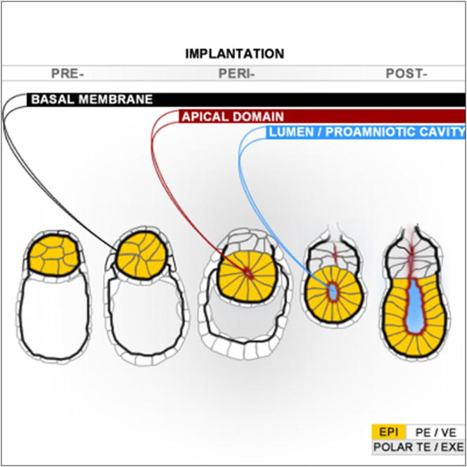
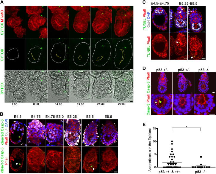
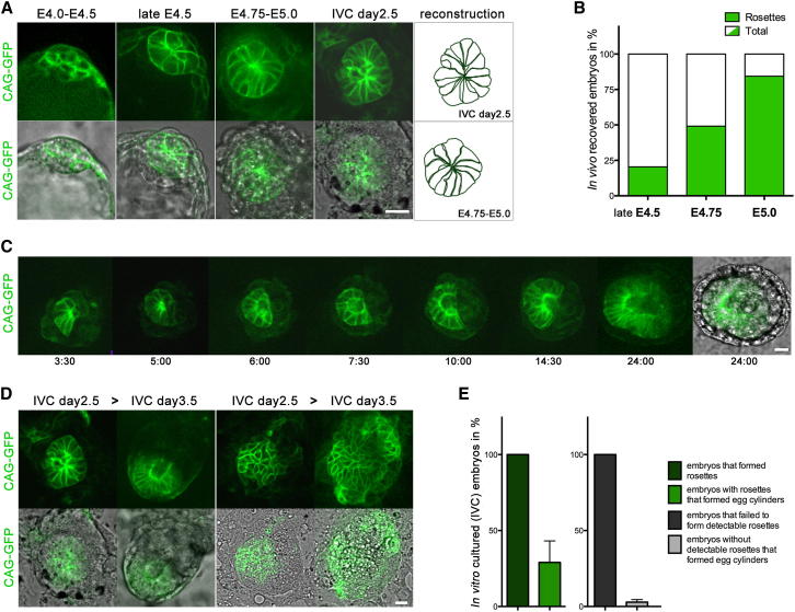
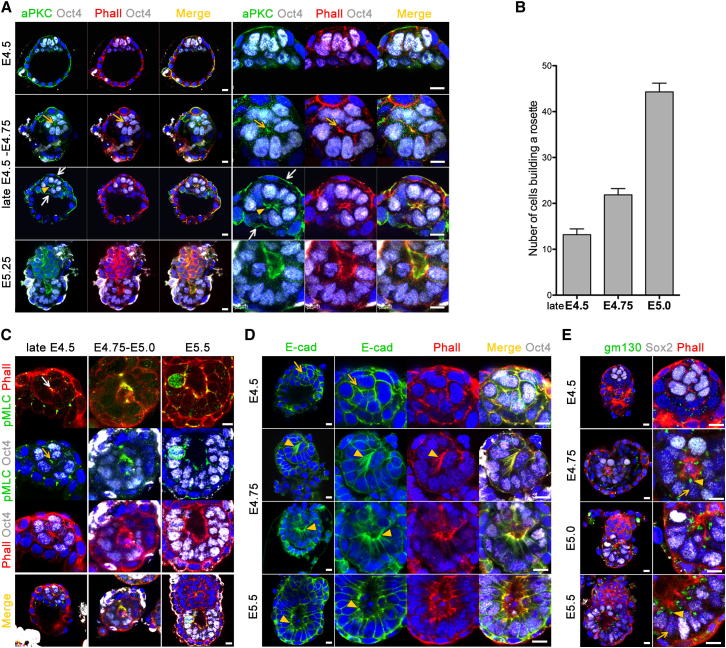
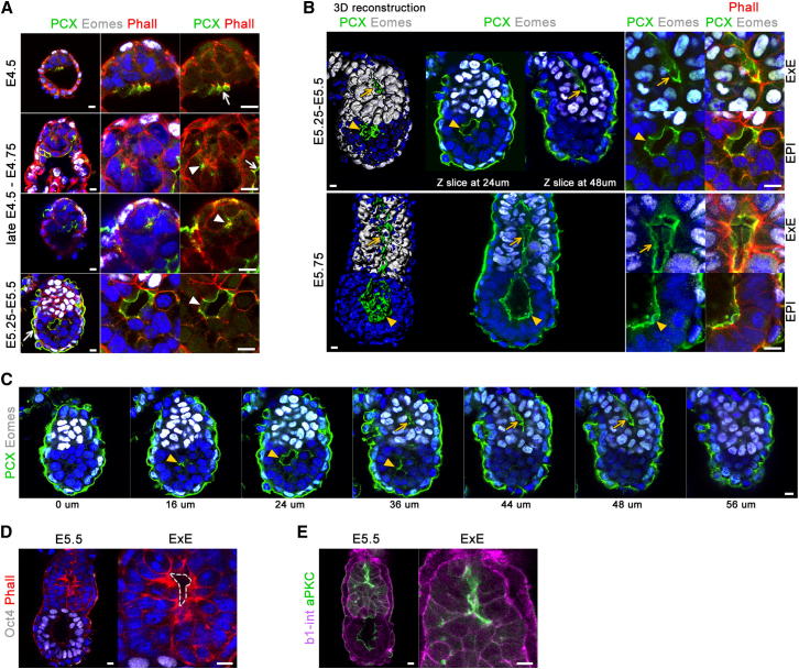
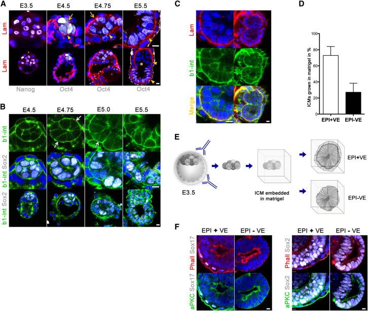
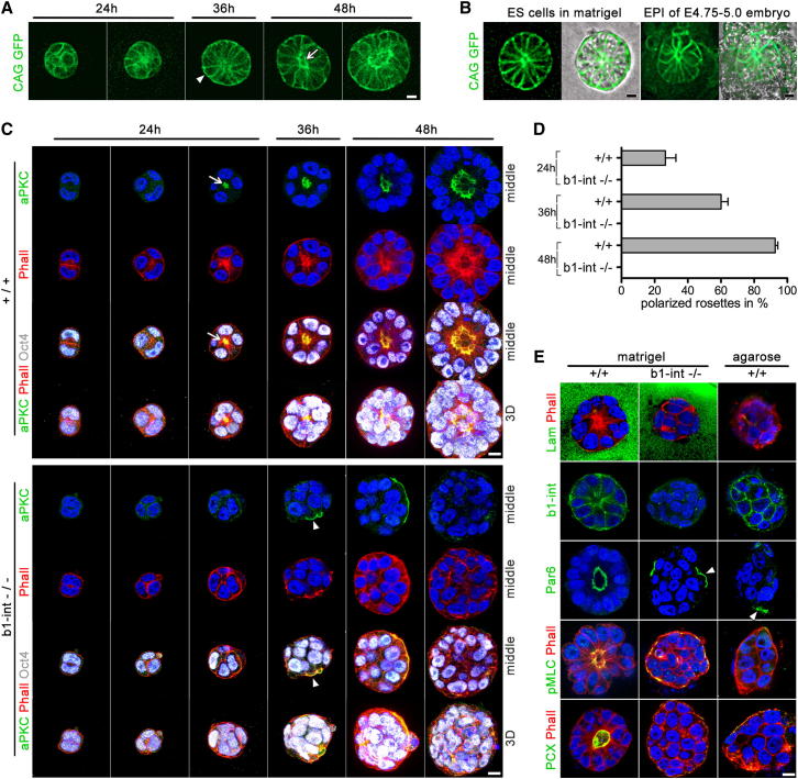
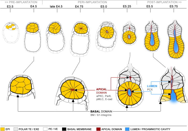
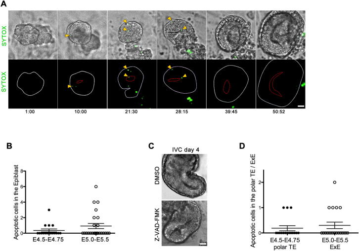
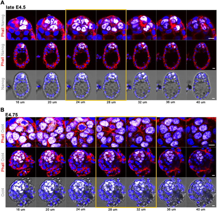
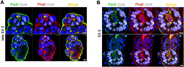
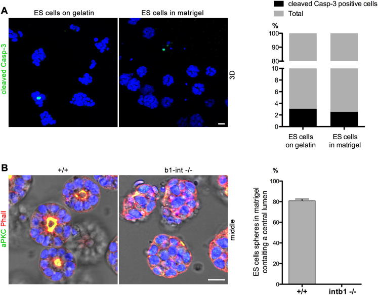
Transformation of pluripotent epiblast cells into a cup-shaped epithelium as the mouse blastocyst implants is a poorly understood and yet key developmental step. Studies of morphogenesis in embryoid bodies led to the current belief that it is programmed cell death that shapes the epiblast. However, by following embryos developing in vivo and in vitro, we demonstrate that not cell death but a previously unknown morphogenetic event transforms the amorphous epiblast into a rosette of polarized cells. This transformation requires basal membrane-stimulated integrin signaling that coordinates polarization of epiblast cells and their apical constriction, a prerequisite for lumenogenesis. We show that basal membrane function can be substituted in vitro by extracellular matrix (ECM) proteins and that ES cells can be induced to form similar polarized rosettes that initiate lumenogenesis. Together, these findings lead to a completely revised model for peri-implantation morphogenesis in which ECM triggers the self-organization of the embryo's stem cells.


Figures

None
Graphical abstract
Figure 1
Figure 1
Cell Death in the Emerging Egg Cylinder (A) Still images of time-lapse recording of MT/MG embryo forming egg cylinder in vitro. Dying cells are marked by SYTOX, a green cell death reporter (IVC embryos n = 23). A yellow line marks the site of the emerging proamniotic cavity. (B) E4.5–E5.5 embryos stained for cleaved caspase-3 (arrowheads). EPI is marked by Oct4 staining (white), F-actin is labeled by phalloidin (red), and the nuclei are counterstained with DAPI (blue). (C) Distribution of dying cells marked by TUNEL (green) at E4.5–E4.75 (n = 11 embryos) and E5.0–E5.5 (n = 27 embryos). EPI is stained for Oct4 (white), phalloidin (red), and DAPI (blue). (D) E5.5 p53 heterozygous (+/−) and KO (−/−) embryos stained for cleaved caspase-3 (green). F-actin is labeled by phalloidin (red), and the nuclei are counterstained with DAPI (blue). (E) Apoptotic cells in the EPI of E5.5–E6.0 embryos derived from p53 heterozygous intercrosses (p53 +/− and +/+ n = 34 embryos, p53 −/− n = 12 embryos). Almost no cleaved caspase-3-positive cells were found in the p53 KO embryos in comparison to WT and heterozygous littermates. p = 0.0411, t test. Error bars represent SEM. Scale bars, 20 μm. See also Figure S1.
Figure 2
Figure 2
The Cells of the Peri-Implantation EPI Organize into a Rosette (A) Organization of the embryonic lineage in CAG-GFP embryos revealed by high membrane GFP expression in the EPI. EPI cells change their shape from round at preimplantation (E4.0–E4.5) to wedge shaped at peri-implantation stages in vivo (late E4.5–E5.0) as well as in vitro (IVC day 2.5). The wedge-shaped cells of the EPI are arranged as a rosette. (B) The amount of embryos with EPI organized as a rosette steadily increases from late E4.5 (20.3%, n = 11/54 embryos), E4.75 (49.1%, n = 28/57 embryos), to E5.0 (84.4%, n = 27/32 embryos). (C) Still images of time-lapse recording of in-vitro-cultured CAG-GFP embryo. Note that a single cavity emerges from the center of the rosette. (D) Representative snapshots of the in vitro development of individual CAG-CAG embryos. On the left, embryo with EPI rosette developed to egg cylinder. The embryo on the right failed to form a rosette and remained poorly organized. (E) In vitro, 28% of embryos that formed rosettes developed to egg cylinders (total embryos n = 36). In contrast, only 4% of embryos without detectable rosettes gave rise to egg cylinders (total embryos n = 75), four independent experiments. Error bars represent SEM. Scale bars, 20 μm. See also Figure S2.
Figure 3
Figure 3
Establishment of Epithelial Polarity in the EPI (A) De novo establishment of the apical domain in EPI cells. E4.5–E5.25 embryos were stained for the apical marker aPKC (green), EPI is stained for Oct4 (white), and F-actin is labeled by phalloidin (red). Low-magnification images are presented in the left, and high magnification of the EPI is in the right. (B) The number of cells building polarized rosettes per developmental stage is steadily increasing. At late E4.5, the average cell number is 13 (n = 11 embryos), at E4.75 it is 22 (n = 35 embryos), and at E5.0 it is 44 (n = 27 embryos). Only cells expressing specific EPI markers such as Oct4, Nanog, or Sox2 were counted. Error bars represent SEM. (C) Myosin II regulatory light-chain phosphorylation revealed by pMLC antibody staining (green). EPI is stained for Oct4 (white) and phalloidin (red). (D) Localization of adherens junctions revealed by E-cad staining (green). EPI is labeled by Oct4 staining (white) and phalloidin (red). (E) Localization of the Golgi apparatus is marked by gm130 antibody staining (green). The embryonic lineage is stained for Sox2 (white) and phalloidin (red). Scale bars, 10 μm. See also Figure S3.
Figure 4
Figure 4
Lumenogenesis in the Peri-Implantation Embryo (A) Expression and localization of podocalyxin (PCX) in early embryos (green). The TE/ExE and the VE are labeled by Eomes (white) and phalloidin (red). (B) 3D reconstructions and optical sections of early egg cylinders stained for PCX (green) and Eomes (white). Higher magnification of the EPI and ExE regions is shown in the right, phalloidin (red) and DAPI (blue). 3D reconstruction was performed using Imaris software. (C) Series of optical sections of E5.25–E5.5 embryo stained for podocalyxin (green) and Eomes (white). The ExE lumenal space is indicated with arrows, and the EPI lumen is indicated with arrowheads. (D) Apical domains marked by concentrated F-actin (phalloidin, red) in ExE cells enclosing a lumen. Oct4 (white) labels the EPI. (E) aPKC antibody staining (green) of apical domains enclosing an intermembranous slit in the EXE. β1-integrin staining is shown in magenta. Nuclei are counterstained with DAPI. Scale bars, 10 μm.
Figure 5
Figure 5
The Basal Membrane Creates a Niche for the Polarizing EPI (A) TE (arrow) and PE (arrowhead) basal membranes stained for laminin (red) wrap the EPI, labeled by Oct4 or Nanog (white). As the egg cylinder emerges, the basal membrane between the ExE and the EPI is no longer maintained (star), but a common VE basal membrane surrounds both EPI and ExE (arrowheads). (B) Expression of β1-integrin in early embryos (green); EPI is stained for Sox2 (white). (C) Colocalization of β1-integrin (green) and laminin (red) at the basal site of the EPI. (D) E3.5 ICMs were isolated by immunosurgery and embedded in matrigel. The majority of the EPI structures contained VE layer (72.8% in average, total ICMs n = 65), but some of the EPIs were in direct contact with the matrigel (27.1% of total ICMs n = 65); four independent experiments. Error bars represent SEM. (E) Schematic representation of the experimental procedure. (F) ICMs isolated by immunosurgery gave rise to polarized EPI structures surrounded by VE or directly contacting the ECM. Apical domains are marked by aPKC (green), VE is stained for Sox17 (white), and EPI is stained for Sox2 (white); phalloidin (red), DAPI (blue). Scale bars, 10 μm.
Figure 6
Figure 6
Self-Organization of ES Cell Spheres Grown in 3D ECM (A) Still images of time-lapse recording of CAG-GFP ES cells’ self-organization and lumenogenesis. (B) CAG-GFP ES cells embedded in matrigel (left) compared to the peri-implantation EPI of E4.75–E5.0 CAG-GFP embryo (right). (C) 3D projections and sections through the center of WT and β1-integrin KO ES cell spheres embedded in matrigel and stained for aPKC (green), Oct4 (white), and phalloidin (red). (D) Formation of polarized rosettes in WT (+/+) versus β1-integrin ko (b1-int −/−) ES cells cultured in matrigel; the rate of self-organization in WT ES cell spheres was examined at 24 hr (26.5% of total spheres n = 82), 36 hr (60% of n = 195), and 48 hr (92.6% of n = 394) by aPKC or Par6 staining in > four independent experiments. β1-integrin KO ES cells failed to properly self-organize at all time points (24 hr total ES cells spheres n = 200; 36 hr n = 123; 48 hr n = 421). Error bars represent SEM. (E) Marker analysis of WT and β1-integrin ko ES cells embedded in matrigel and WT ES cells embedded in agarose. Scale bars, 10 μm. See also Figure S4.
Figure 7
Figure 7
A Model for the Sequence of the Morphogenic Events that Drive the Peri-Implantation Development The pre-implantation blastocyst is comprised of unpolarized EPI cells and polarized epithelia of the TE and PE (E4.5). The cells of the extraembryonic lineages secrete ECM proteins that assemble a basal membrane. The basal membrane is in direct contact on one side with the basal domain of the TE or PE cells and on the other with the EPI cells. Thereby, the basal membrane wraps around the embryonic lineage and creates a niche for its maturation during the implantation period (late E4.5–E5.25). β1-integrin receptors on the interface between the cells and the ECM enable the EPI to sense the components of the basal membrane. The ECM proteins provide polarization cues that orient the establishment of a basal-apical axis of the EPI cells. The cell membrane in direct contact with the basal membrane in the EPI periphery adopts basal identity, whereas the opposite membrane domain in the EPI center acquires apical features. Accumulation of apical determinants such as components of the Par complex establish the apical domain de novo. The cells change their shape as a result of actomyosin constriction, coupled to apical localization of adherens junctions. These radially orientated wedge-shaped cells form a rosette-like structure (E4.75–E5.0). A small lumenal space appears in the center of the rosette. The lumen is formed by hollowing, as a result of a charge repulsion of apical membranes coated by anti-adhesive glycoproteins such as podocalyxin (E5.0–E5.25). The lumen broadens as the egg cylinder elongates. Exocytosis and pumping could enhance fluid filling and could contribute to its enlargement. Similar processes of polarization and membrane repulsion generate small slits between the apical domains of ExE cells (E5.5). At that time, the EPI and the ExE are in direct contact, as the basal membrane initially covering the proximal cells of the EPI is no longer maintained. The basal membrane resembles a basket in which the basal sites of the EPI cells are anchored through integrins. Thus, the basal membrane might act as a mold to determine the global shape of the EPI, transforming it from a symmetric hollowed sphere to a cup. The lumen of the EPI cup enlarges and, together with ExE intermembranous space, forms the mature proamniotic cavity. At this point, the egg cylinder formation is complete (E5.5–E5.75).
Figure S1
Figure S1
Cell Death during the Peri-Implantation Stages of Development, Related to Figure 1 (A) Still images of time-lapse recording of emerging egg-cylinder in vitro. Dying cells are marked by SYTOX. A red line marks the site of the proamniotic cavity. (B) Quantification of the Cleaved Caspase-3 positive cells in the EPI of E4.5-75 (n = 17 embryos) and E5.0-5.5 embryos (n = 26 embryos). Error bars represent SEM. (C) IVC embryos formed proamniotic cavity in the presence of 20 μM Z-DEVD FMK (n = 26 embryos), similar to DMSO treated control embryos (n = 21). Scale bar = 45 μm. (D) Quantification of the Cleaved Caspase-3 positive cells in the polar TE of E4.5-75 (n = 16 embryos) and the ExE of E5.0-5.5 embryos (n = 20 embryos). Error bars represent SEM.
Figure S2
Figure S2
EPI Organizes as a Rosette in Peri-Implantation Embryos with Blastocyst Morphology, Related to Figure 2 (A and B) Optical Z-section series of immunofluorescence confocal images of late E4.5 (A) and E4.75 embryos (B). Nanog or Oct4 positive EPI cells (white) are organized as a rosette (yellow lined panels). F-actin is labeled by phalloidin (red), the nuclei are counterstained with DAPI (blue). Scale bars = 10 μm.
Figure S3
Figure S3
Polarization of the EPI during the Peri-Implantation Stages of Development, Related to Figure 3 (A and B) (A) Par6 staining (green) reveals the establishment of the apical domain in EPI cells of the late E4.5 embryo, prior to lumen formation at E5.5 (B); EPI is labeled by Oct4 (white), phalloidin (red), nuclei are counterstained with DAPI. Scale bars = 10 μm.
Figure S4
Figure S4
Apoptosis and Formation of a Central Lumen in ES Cell Spheres Grown in 3D ECM, Related to Figure 6 (A) Apoptosis in wild-type ES cell colonies grown on gelatin-coated plates and ES cell spheres cultured in matrigel for 48h. Apoptotic cells are stained for cleaved Caspase-3 (green) and the nuclei are counterstained with DAPI (blue). The proportion of cleaved Caspase-3 positive cells is similar between cells cultured on gelatin-coated plates (3%, total n = 1186 cells) versus cells cultured in 3D matrigel gels (2.5%, total n = 1012 cells). (B) Wild-type (+/+) and β1-integrin ko (b1-int −/−) ES cells cultured in matrigel and stained for aPKC (green) and phalloidin (red). 80.7% of the wild-type ES cell spheres contained a central lumen (total n = 225 ES cell spheres) in contrast to the β1-integrin ko spheres that remained poorly organized (n = 169). Scale bars = 20 μm. Error bars represent SEM.

Similar articles

Cited by

References

    1. Arman E., Haffner-Krausz R., Chen Y., Heath J.K., Lonai P. Targeted disruption of fibroblast growth factor (FGF) receptor 2 suggests a role for FGF signaling in pregastrulation mammalian development. Proc. Natl. Acad. Sci. USA. 1998;95:5082–5087. - PMC - PubMed
    1. Bedzhov I., Alotaibi H., Basilicata M.F., Ahlborn K., Liszewska E., Brabletz T., Stemmler M.P. Adhesion, but not a specific cadherin code, is indispensable for ES cell and induced pluripotency. Stem Cell Res. (Amst.) 2013;11:1250–1263. - PubMed
    1. Bryant D.M., Mostov K.E. From cells to organs: building polarized tissue. Nat. Rev. Mol. Cell Biol. 2008;9:887–901. - PMC - PubMed
    1. Chazaud C., Yamanaka Y., Pawson T., Rossant J. Early lineage segregation between epiblast and primitive endoderm in mouse blastocysts through the Grb2-MAPK pathway. Dev. Cell. 2006;10:615–624. - PubMed
    1. Chen F., Ma L., Parrini M.C., Mao X., Lopez M., Wu C., Marks P.W., Davidson L., Kwiatkowski D.J., Kirchhausen T. Cdc42 is required for PIP(2)-induced actin polymerization and early development but not for cell viability. Curr. Biol. 2000;10:758–765. - PubMed

Supplemental References

    1. Bedzhov I., Liszewska E., Kanzler B., Stemmler M.P. Igf1r signaling is indispensable for preimplantation development and is activated via a novel function of E-cadherin. PLoS Genet. 2012;8:e1002609. - PMC - PubMed
    1. Vestweber D., Kemler R. Rabbit antiserum against a purified surface glycoprotein decompacts mouse preimplantation embryos and reacts with specific adult tissues. Exp. Cell Res. 1984;152:169–178. - PubMed