

Inactivated human platelet lysate with psoralen: a new perspective for mesenchymal stromal cell production in Good Manufacturing Practice conditions
- PMID: 24529555
- PMCID: PMC7185570
- DOI: 10.1016/j.jcyt.2013.12.008
Inactivated human platelet lysate with psoralen: a new perspective for mesenchymal stromal cell production in Good Manufacturing Practice conditions
Abstract
Background aims: Mesenchymal stromal cells (MSC) are ideal candidates for regenerative and immunomodulatory therapies. The use of xenogeneic protein-free Good Manufacturing Practice-compliant growth media is a prerequisite for clinical MSC isolation and expansion. Human platelet lysate (HPL) has been efficiently implemented into MSC clinical manufacturing as a substitute for fetal bovine serum (FBS). Because the use of human-derived blood materials alleviates immunologic risks but not the transmission of blood-borne viruses, the aim of our study was to test an even safer alternative than HPL to FBS: HPL subjected to pathogen inactivation by psoralen (iHPL).
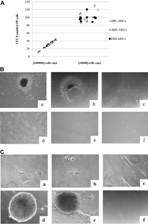
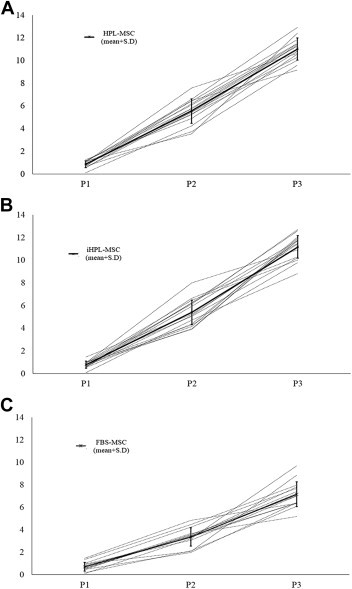
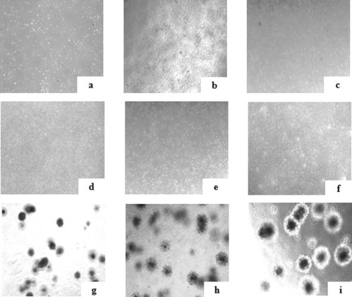
Methods: Bone marrow samples were plated and expanded in α-minimum essential medium with 10% of three culture supplements: HPL, iHPL and FBS, at the same time. MSC morphology, growth and immunophenotype were analyzed at each passage. Karyotype, tumorigenicity and sterility were analyzed at the third passage. Statistical analyses were performed.
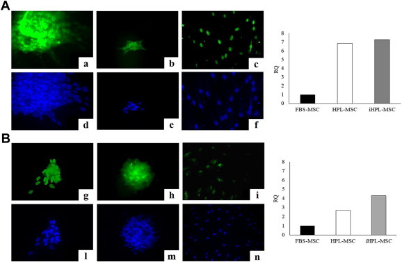
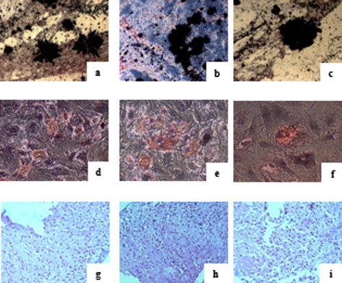
Results: The MSCs cultivated in the three different culture conditions showed no significant differences in terms of fibroblast colony-forming unit number, immunophenotype or in their multipotent capacity. Conversely, the HPL/iHPL-MSCs were smaller, more numerous, had a higher proliferative potential and showed a higher Oct-3/4 and NANOG protein expression than did FBS-MSCs. Although HPL/iHPL-MSCs exhibit characteristics that may be attributable to a higher primitive stemness than FBS-MSCs, no tumorigenic mutations or karyotype modifications were observed.
Conclusions: We demonstrated that iHPL is safer than HPL and represents a good, Good Manufacturing Practice-compliant alternative to FBS for MSC clinical production that is even more advantageous in terms of cellular growth and stemness.
Keywords: Good Manufacturing Practice; human platelet lysate; inactivation; mesenchymal stromal cells; psoralen.
Copyright © 2014 International Society for Cellular Therapy. Published by Elsevier Inc. All rights reserved.
Figures
Comment in
-
Finessing the manufacture of mesenchymal stromal cells.Cytotherapy. 2014 Jun;16(6):711-2. doi: 10.1016/j.jcyt.2014.04.001. Cytotherapy. 2014. PMID: 24801376 No abstract available.
References
-
- Pittenger M.F., Mackay A.M., Beck S.C., Jaiswal R.K., Douglas R., Mosca J.D. Multilineage potential of adult human mesenchymal stem cells. Science. 1999;284:143–147. - PubMed
-
- Mareschi K., Biasin E., Piacibello W., Aglietta M., Madon E., Fagioli F. Isolation of human mesenchymal stem cells: bone marrow versus umbilical cord blood. Haematologica. 2001;86:1099–1100. - PubMed
-
- Aggarwal S., Pittenger M.F. Human mesenchymal stem cells modulate allogeneic immune cell responses. Blood. 2005;105:1815–1822. - PubMed
-
- Mazzini L., Mareschi K., Ferrero I., Vassallo E., Oliveri G., Boccaletti R. Autologous mesenchymal stem cells: clinical applications in amyotrophic lateral sclerosis. Neurol Res. 2006;28:523–526. - PubMed
MeSH terms
Substances
LinkOut - more resources
Full Text Sources
Other Literature Sources
Research Materials
