. 2014 Mar 6;14(3):323-328.
doi: 10.1016/j.stem.2014.01.018. Epub 2014 Feb 13.

TALEN-mediated gene mutagenesis in rhesus and cynomolgus monkeys

Affiliations

TALEN-mediated gene mutagenesis in rhesus and cynomolgus monkeys

Hailiang Liu et al. Cell Stem Cell. .

Abstract

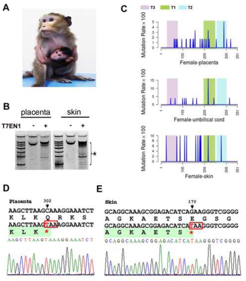
Recent advances in gene editing technology have introduced the potential for application of mutagenesis approaches in nonhuman primates to model human development and disease. Here we report successful TALEN-mediated mutagenesis of an X-linked, Rett syndrome (RTT) gene, methyl-CpG binding protein 2 (MECP2), in both rhesus and cynomolgus monkeys. Microinjection of MECP2-targeting TALEN plasmids into rhesus and cynomolgus zygotes leads to effective gene editing of MECP2 with no detected off-target mutagenesis. Male rhesus (2) and cynomolgous (1) fetuses carrying MECP2 mutations in various tissues including testes were miscarried during midgestation, consistent with RTT-linked male embryonic lethality in humans. One live delivery of a female cynomolgus monkey occurred after 162 days of gestation, with abundant MECP2 mutations in peripheral tissues. We conclude that TALEN-mediated mutagenesis can be an effective tool for genetic modeling of human disease in nonhuman primates.


Figures

Figure 1
Figure 1. MECP2 mutant monkey fetuses generated by TALEN (see also Figure S1 in the supplement)
(A) Schematic of TALEN-targeting sites within monkey MECP2 locus exon 3. The first nucleotides of exon 3 is numbered as 1, the last nucleotides of exon 3 is number 351. TALEN-targeting sequences are labeled as T1, T2, T3 (L, R). (B) Summary of embryo transfer (ET) after TALEN injection in both rhesus and cynomolgous monkeys (#, embryos transferred between 2-cell and blastocyst stage; a, including 1 singleton and 1 twin; b, miscarriage happened on the 30th and 63rd/64th days after ET; c, one female cynomolgus was born alive after 162 days of gestation; d, one male cynomolus fetus was miscarried on the 92nd day of gestation). (C) Images and gender identification of the two aborted Rhesus monkey fetuses. (D) Paternal and maternal DNAs were subjected to Sanger sequencing and T7EN1 cleavage assays. Since the paternal allele is homogenous, T7EN1 is incapable to cut. The maternal alleles are heterogeneous (with one 217C allele and one 217T allele), thus in T7En1 cleavage assay, positive digestion was observed (two arrows point at cleaved products). (E) Positive T7EN1 cleavage results from the miscarried monkey fetuses’ tail, brain, and testis tissues, indicative of presence of mutations at the targeted exon (cleaved products are indicated by the bracket with a star). (F) All detected point mutations were plotted based on its position and mutation rate in the entire 351bp exon. Mutation distribution plots were displayed for tail, brain and testes of the two miscarried fetuses. (G) Detrimental mutations (leading to early truncations or in-dels) detected in brain and tails of the fetuses. (H) Mutation rates calculated from Sanger sequencing of MECP2 exon 3. From total clones sequenced, we counted perfectly matched sequences with reference and sequences with at least one mismatch. When the percentage of perfectly matched sequences were calculated, we simultaneously acquired the mutation rate in each tissues sequenced (see also Table S1 and Figure S2 in the supplement).
Figure 2
Figure 2. Generation of TALEN-targeted live cynomolgus female monkey
(A) Images of three-month-old MECP2-mutant cynomolgus with her mother. (B) T7EN1 cleavage analysis of the placenta and skin tissue of the female cynomolgus baby born alive, indicative of presence of mutations by cleaved products (bracket with a star). (C) Mutation distributions along the targeted exon 3 based on Sanger sequencing in placenta, skin, and umbilical cord. (D-E) Sequencing traits of nonsense mutations detected in placenta and skins of the cynomolgus female baby.

References

    1. Amir RE, Van den Veyver IB, Wan M, Tran CQ, Francke U, Zoghbi HY. Rett syndrome is caused by mutations in X-linked MECP2, encoding methyl-CpG-binding protein 2. Nat. Genet. 1999;23:185–188. - PubMed
    1. Chan AW, Chong KY, Martinovich C, Simerly C, Schatten G. Transgenic monkeys produced by retroviral gene transfer into mature oocytes. Science. 2001;291:309–312. - PubMed
    1. Chen Y, Niu Y, Ji W. Transgenic nonhuman primate models for human diseases: approaches and contributing factors. J. Genet. Genomics. 2012a;39:247–251. - PubMed
    1. Chen Y, Niu Y, Yang S, He X, Ji S, Si W, Tang X, Xie Y, Wang H, Lu Y, Zhou Q, Ji W. The available time window for embryo transfer in the rhesus monkey (Macaca mulatta). Am. J. Primatol. 2012b;74:165–173. - PubMed
    1. Cho SW, Kim S, Kim JM, Kim JS. Targeted genome engineering in human cells with the Cas9 RNA-guided endonuclease. Nat. Biotechnol. 2013;31:230–232. - PubMed

Publication types