. 2014 Mar 6;94(3):453-61.
doi: 10.1016/j.ajhg.2014.01.006. Epub 2014 Feb 13.

Mitochondrial carbonic anhydrase VA deficiency resulting from CA5A alterations presents with hyperammonemia in early childhood

Affiliations

Mitochondrial carbonic anhydrase VA deficiency resulting from CA5A alterations presents with hyperammonemia in early childhood

Clara D van Karnebeek et al. Am J Hum Genet. .

Abstract

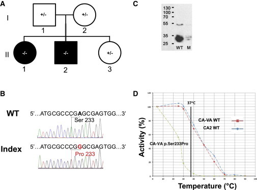
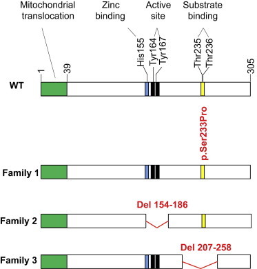
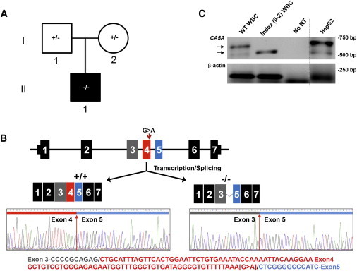
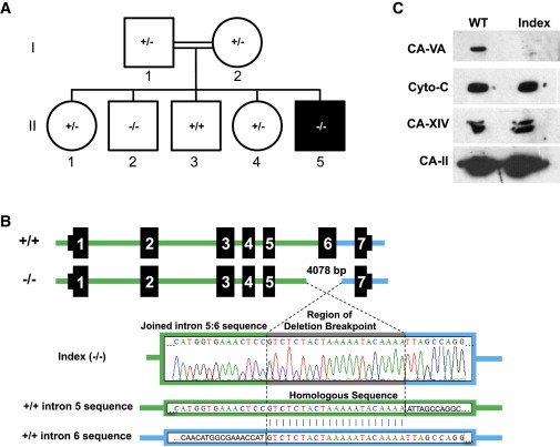
Four children in three unrelated families (one consanguineous) presented with lethargy, hyperlactatemia, and hyperammonemia of unexplained origin during the neonatal period and early childhood. We identified and validated three different CA5A alterations, including a homozygous missense mutation (c.697T>C) in two siblings, a homozygous splice site mutation (c.555G>A) leading to skipping of exon 4, and a homozygous 4 kb deletion of exon 6. The deleterious nature of the homozygous mutation c.697T>C (p.Ser233Pro) was demonstrated by reduced enzymatic activity and increased temperature sensitivity. Carbonic anhydrase VA (CA-VA) was absent in liver in the child with the homozygous exon 6 deletion. The metabolite profiles in the affected individuals fit CA-VA deficiency, showing evidence of impaired provision of bicarbonate to the four enzymes that participate in key pathways in intermediary metabolism: carbamoylphosphate synthetase 1 (urea cycle), pyruvate carboxylase (anaplerosis, gluconeogenesis), propionyl-CoA carboxylase, and 3-methylcrotonyl-CoA carboxylase (branched chain amino acids catabolism). In the three children who were administered carglumic acid, hyperammonemia resolved. CA-VA deficiency should therefore be added to urea cycle defects, organic acidurias, and pyruvate carboxylase deficiency as a treatable condition in the differential diagnosis of hyperammonemia in the neonate and young child.


Figures

Figure 1
Figure 1
Family 1 with p.Ser233Pro Missense Variant (A) Pedigree (black fill indicates clinically affected individuals, II-1 and II-2). (B) Sanger sequence of CA5A from index (II-1) and control (wild-type sequence; WT) subjects; the variant nucleotide position and the corresponding codon alteration (p.Ser233Pro) are indicated. (C) Immunoblot analyses by SDS-PAGE (ImageJ software) of WT and p.Ser233Pro (mutant; M) CA-VA protein levels in COS-7 cell lysates; the molecular weights (kDa) of protein standards are indicated on the left. Normal and mutant (c.697T>C) CA5A cDNAs, including the full mitochondrial targeting sequences, were synthesized via the NCBI reference sequence (NM_001739.1) by Genscript. The cDNAs were cloned into pUC57 at XhoI and BglII sites and verified by Sanger sequencing. Subsequently, the cDNA inserts were subcloned via the same restriction sites into the pCXN mammalian expression vector. COS-7 cells (ATCC CRL-1651) were transfected with wild-type or mutant CA5A plasmids with Lipofectamine 2000 (Invitrogen) according to the manufacturer’s protocol, as previously described. A β-glucuronidase expression plasmid was cotransfected as a marker of transfection efficiency. COS-7 cells were harvested 48–72 hr after transfection in lysis buffer and then lysed by sonication on ice. (D) Thermal stability profiles for WT (red) and p.Ser233Pro mutant (green) CA-VA enzymes. Carbonic anhydrase II (CA2; blue) was used as a control.
Figure 2
Figure 2
Effect of Genetic Variants Identified in CA-VA Shown is a schematic diagram of the 305 amino acid wild-type (WT) CA-VA. Residues 1–39 encode a mitochondrial translocation signal (green). Homology predictions indicate that histidine 155 binds a zinc ion (blue), tyrosines 164 and 167 are active site residues (black), and threonines 235 and 236 comprise a substrate-binding region (yellow). Shown in red are the deduced CA-VA variants identified in this study. The index and affected brother of family 1 has a nonsynonymous Ser to Pro mutation at residue 233, adjacent to the substrate-binding region. The index of family 2 has a deletion of residues 154–186 (exon 4), thereby missing the metal-binding and active-site residues. The index of family 3 has a deletion of residues 207–258 (exon 6; the substrate-binding region), which results in absent protein.
Figure 3
Figure 3
Family 2 with Exon 4 Splice Deletion (A) Pedigree (black fill indicates clinically affected individual). (B) Sanger sequencing of RT-PCR products generated in (B) with exons denoted by colors. Top: The CA5A structure (not to scale) and a schematic of the observed CA5A transcripts produced in a control subject and the index. Bottom: Sanger sequence of transcripts at exon 4 boundary in a control subject (+/+) and the index (−/−), along with WT CA5A cDNA sequence, color-coded as in the top panel. (C) RT-PCR of CA5A mRNA from white blood cells (WBCs) or cultured liver cells (HepG2). Arrows indicate the products of differing size amplified from control subject (WT sequence) and index (II-1) WBCs. As controls, reverse transcriptase was omitted from the reaction (no RT) and a control gene (β-actin) was amplified in separate lane on a different cell type (denoted by the line).
Figure 4
Figure 4
Family 3 with Exon 6 Deletion (A) Pedigree (black fill indicates clinically affected individual, II-5). (B) Top: Schematic representation of the 4,078 bp deletion that encompasses exon 6 of CA5A. Bottom: Sanger sequencing of PCR products generated from genomic DNA of the index (II-5) with a 21 bp repeated sequence (gray) at the breakpoint found in both intron 5 (green) and intron 6 (blue). (C) Immunoblot analyses of control subject (WT) and index (II-5) liver homogenates. Samples were probed for CA-VA, cytochrome c (Cyto-c), carbonic anhydrase XIV (CA 14), and carbonic anhydrase II (CA 2) via specific antibodies.
Figure 5
Figure 5
Biochemical Pathways Affected by CA-VA Deficiency CA-VA deficiency is indicated by the horizontal red bar (bottom right). Dotted lines link CA-VA-mediated bicarbonate production and intramitochondrial donation to the bicarbonate-dependent carboxylases; the affected enzyme deficiencies are denoted with red circles. Enzymes: CPS1, carbamoylphosphate synthetase I; PC, pyruvate carboxylase; PCC, propionyl CoA carboxylase; 3MCC, 3-methylcrotonyl CoA carboxylase. Metabolites: SUC, succinyl CoA; αKG, α-ketoglutarate; OAA, oxaloacetate; Glu, glutamate; Gln, glutamine; Sac, saccharopine; Lys, lysine; ASP, aspartate; PEP, phosphoenolpyruvate; PYR, pyruvate; LAC, lactate; AcCoA, acetyl CoA; AS, argininosuccinate; 3MGCoA, 3-methylglutaconyl CoA; 3MCCoA, 3-methylcrotonyl CoA.

References

    1. Häberle J. Clinical and biochemical aspects of primary and secondary hyperammonemic disorders. Arch. Biochem. Biophys. 2013;536:101–108. - PubMed
    1. Daniotti M., la Marca G., Fiorini P., Filippi L. New developments in the treatment of hyperammonemia: emerging use of carglumic acid. Int. J. Gen. Med. 2011;4:21–28. - PMC - PubMed
    1. Nagao Y., Batanian J.R., Clemente M.F., Sly W.S. Genomic organization of the human gene (CA5) and pseudogene for mitochondrial carbonic anhydrase V and their localization to chromosomes 16q and 16p. Genomics. 1995;28:477–484. - PubMed
    1. Krebs J.F., Fierke C.A. Determinants of catalytic activity and stability of carbonic anhydrase II as revealed by random mutagenesis. J. Biol. Chem. 1993;268:948–954. - PubMed
    1. Andresen B.S., Dobrowolski S.F., O’Reilly L., Muenzer J., McCandless S.E., Frazier D.M., Udvari S., Bross P., Knudsen I., Banas R. Medium-chain acyl-CoA dehydrogenase (MCAD) mutations identified by MS/MS-based prospective screening of newborns differ from those observed in patients with clinical symptoms: identification and characterization of a new, prevalent mutation that results in mild MCAD deficiency. Am. J. Hum. Genet. 2001;68:1408–1418. - PMC - PubMed

Publication types

MeSH terms

Substances