

Antibacterial activity of rifamycins for M. smegmatis with comparison of oxidation and binding to tear lipocalin
- PMID: 24530503
- PMCID: PMC3992280
- DOI: 10.1016/j.bbapap.2014.02.001
Antibacterial activity of rifamycins for M. smegmatis with comparison of oxidation and binding to tear lipocalin
Abstract
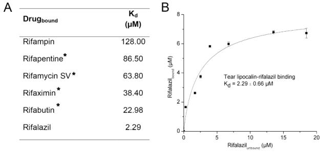
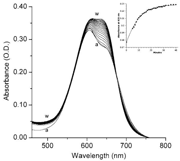
A mutant of Mycobacterium smegmatis is a potential class I model substitute for Mycobacterium tuberculosis. Because not all of the rifamycins have been tested in this organism, we determined bactericidal profiles for the 6 major rifamycin derivatives. The profiles closely mirrored those established for M. tuberculosis. Rifalazil was confirmed to be the most potent rifamycin. Because the tuberculous granuloma presents a harshly oxidizing environment we explored the effects of oxidation on rifamycins. Mass spectrometry confirmed that three of the six major rifamycins showed autoxidation in the presence of trace metals. Oxidation could be monitored by distinctive changes including isosbestic points in the ultraviolet-visible spectrum. Oxidation of rifamycins abrogated anti-mycobacterial activity in M. smegmatis. Protection from autoxidation was conferred by binding susceptible rifamycins to tear lipocalin, a promiscuous lipophilic protein. Rifalazil was not susceptible to autoxidation but was insoluble in aqueous solution. Solubility was enhanced when complexed to tear lipocalin and was accompanied by a spectral red shift. The positive solvatochromism was consistent with robust molecular interaction and binding. Other rifamycins also formed a complex with lipocalin, albeit to a lesser extent. Protection from oxidation and enhancement of solubility with protein binding may have implications for delivery of select rifamycin derivatives.
Keywords: Lipocalin-1; Mycobacteria smegmatis; Oxidation; Rifabutin (PubChem CID: 6323490); Rifalazil; Rifalazil (PubChem CID: 6540558); Rifampin (PubChem CID: 24871024); Rifamycin; Rifamycin S (PubChem CID: 6436726); Rifamycin SV (PubChem CID: 6324616); Rifapentine (PubChem CID: 6323497); Rifaximin (PubChem CID: 6436173); Tear lipocalin.
Copyright © 2014 Elsevier B.V. All rights reserved.
Figures
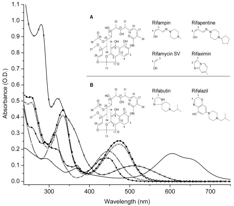

 ), rifapentine (15 μM) (
 ), rifamycin SV (15 μM) (
 ), rifalazil (15 μM) (————), rifaximin (15 μM) (
 ), and rifabutin (30 μM) (
 ). Inset (A, B): Base chemical structures of all six rifamycin drugs (left) and unique groups (right). The hydroxyls at positions 1 and 4 may be oxidized to form the hydroquinone. The sole hydroxyl of the benzoxanino moiety is a candidate for oxidation on rifalazil. Hydroxyls are not available at position 1 and 4 for autoxidation for rifabutin or rifalazil.


Similar articles
-
In vitro activity of new rifamycins against rifampicin-resistant M. tuberculosis and MAIS-complex mycobacteria.Tubercle. 1987 Sep;68(3):177-82. doi: 10.1016/0041-3879(87)90053-5. Tubercle. 1987. PMID: 2834842
-
Novel C-3-(N-alkyl-aryl)-aminomethyl rifamycin SV derivatives exhibit activity against rifampicin-resistant Mycobacterium tuberculosis RpoBS522L strain and display a different binding mode at the RNAP β-subunit site compared to rifampicin.Eur J Med Chem. 2021 Dec 5;225:113734. doi: 10.1016/j.ejmech.2021.113734. Epub 2021 Aug 8. Eur J Med Chem. 2021. PMID: 34418786
-
Rifamycin inhibition of WT and Rif-resistant Mycobacterium tuberculosis and Escherichia coli RNA polymerases in vitro.Tuberculosis (Edinb). 2011 Sep;91(5):361-9. doi: 10.1016/j.tube.2011.05.002. Epub 2011 Jun 24. Tuberculosis (Edinb). 2011. PMID: 21704562
-
Rifamycins--obstacles and opportunities.Tuberculosis (Edinb). 2010 Mar;90(2):94-118. doi: 10.1016/j.tube.2010.02.001. Epub 2010 Mar 16. Tuberculosis (Edinb). 2010. PMID: 20236863 Review.
-
Prospects for development of new antimycobacterial drugs.J Infect Chemother. 2000 Mar;6(1):8-20. doi: 10.1007/s101560050043. J Infect Chemother. 2000. PMID: 11810525 Review.
Cited by
-
Restoration of structural stability and ligand binding after removal of the conserved disulfide bond in tear lipocalin.Biochem Biophys Res Commun. 2014 Oct 3;452(4):1004-8. doi: 10.1016/j.bbrc.2014.09.029. Epub 2014 Sep 16. Biochem Biophys Res Commun. 2014. PMID: 25223802 Free PMC article.
-
Drug Susceptibility Testing of 31 Antimicrobial Agents on Rapidly Growing Mycobacteria Isolates from China.Biomed Res Int. 2015;2015:419392. doi: 10.1155/2015/419392. Epub 2015 Aug 13. Biomed Res Int. 2015. PMID: 26351633 Free PMC article.
-
Ligand binding studies by high speed centrifugal precipitation and linear spectral summation using ultraviolet-visible absorption spectroscopy.MethodsX. 2018 Apr 17;5:345-351. doi: 10.1016/j.mex.2018.04.007. eCollection 2018. MethodsX. 2018. PMID: 30050754 Free PMC article.
-
Tear Lipocalin and Lipocalin-Interacting Membrane Receptor.Front Physiol. 2021 Aug 19;12:684211. doi: 10.3389/fphys.2021.684211. eCollection 2021. Front Physiol. 2021. PMID: 34489718 Free PMC article.
-
Repositioning rifamycins for Mycobacterium abscessus lung disease.Expert Opin Drug Discov. 2019 Sep;14(9):867-878. doi: 10.1080/17460441.2019.1629414. Epub 2019 Jun 14. Expert Opin Drug Discov. 2019. PMID: 31195849 Free PMC article. Review.
References
-
- WHO global tuberculosis control report 2010 Summary. Cent Eur J Public Health. 2010;18:237. - PubMed
-
- Aristoff PA, Garcia GA, Kirchhoff PD, Hollis Showalter HD. Rifamycins--obstacles and opportunities. Tuberculosis (Edinb) 2010;90:94–118. - PubMed
-
- Sayada C. Re: Review article titled, “Rifamycins - Obstacles and opportunities” by Paul A. Aristoff, George A. Garcia, Paul D. Kirchhoff, H.D. Hollis Showalter. Tuberculosis. 2010;90(2):94–118. Tuberculosis (Edinb) 2010;90:326; author reply -7. - PubMed
-
- Snapper SB, Melton RE, Mustafa S, Kieser T, Jacobs WR., Jr. Isolation and characterization of efficient plasmid transformation mutants of Mycobacterium smegmatis. Mol Microbiol. 1990;4:1911–9. - PubMed
Publication types
MeSH terms
Substances
Grants and funding
LinkOut - more resources
Full Text Sources
Other Literature Sources
