. 2014 Jun;10(6):2582-90.
doi: 10.1016/j.actbio.2014.01.032. Epub 2014 Feb 12.

Interfibrillar shear stress is the loading mechanism of collagen fibrils in tendon

Affiliations

Interfibrillar shear stress is the loading mechanism of collagen fibrils in tendon

Spencer E Szczesny et al. Acta Biomater. 2014 Jun.

Abstract

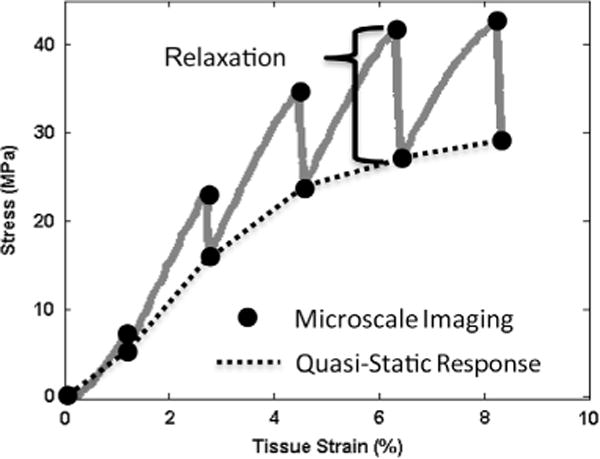
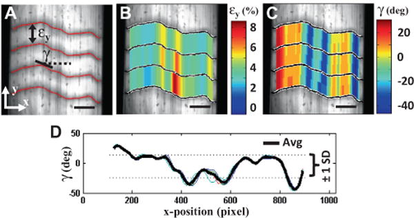
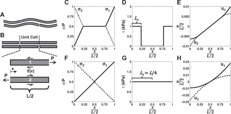
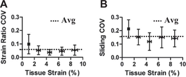
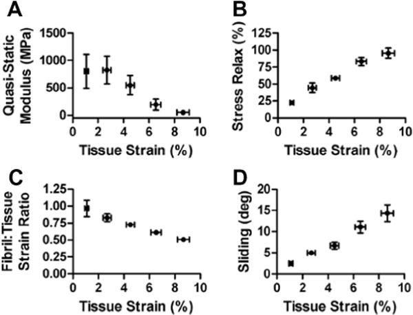
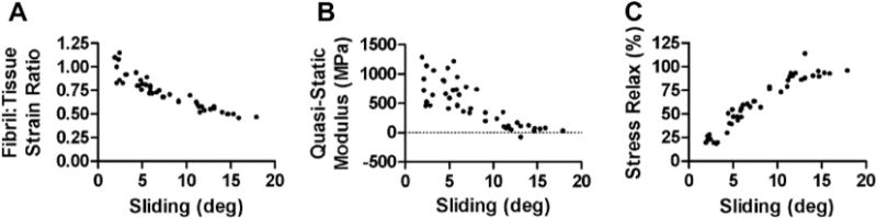
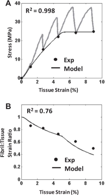
Despite the critical role tendons play in transmitting loads throughout the musculoskeletal system, little is known about the microstructural mechanisms underlying their mechanical function. Of particular interest is whether collagen fibrils in tendon fascicles bear load independently or if load is transferred between fibrils through interfibrillar shear forces. We conducted multiscale experimental testing and developed a microstructural shear lag model to explicitly test whether interfibrillar shear load transfer is indeed the fibrillar loading mechanism in tendon. Experimental correlations between fascicle macroscale mechanics and microscale interfibrillar sliding suggest that fibrils are discontinuous and share load. Moreover, for the first time, we demonstrate that a shear lag model can replicate the fascicle macroscale mechanics as well as predict the microscale fibrillar deformations. Since interfibrillar shear stress is the fundamental loading mechanism assumed in the model, this result provides strong evidence that load is transferred between fibrils in tendon and possibly other aligned collagenous tissues. Conclusively establishing this fibrillar loading mechanism and identifying the involved structural components should help develop repair strategies for tissue degeneration and guide the design of tissue engineered replacements.

Keywords: Fibril sliding; Interfibrillar shear stress; Multiscale testing; Shear lag model; Tendon.


Figures

Fig. 1
Fig. 1
Collagenous hierarchical structure in tendon (adapted from Kastelic et al. [1]). Collagen molecules aggregate into extracellular structures called fibrils, which are the primary tensile load-bearing elements in connective tissues. In tendon, fibrils are highly aligned and form dense macroscopic subunits called fascicles. Also contained within each fascicle are cells whose extended processes loosely separate the fibrils into local partitions called fibers [2,3]. (A, B) Electron micrographs of fibrils and fascicles showing detailed views of corresponding regions highlighted in the hierarchical structure. Micrograph (B) is reprinted with permission from Rowe [2]. Scale bars: (A) 500 nm, (B) 100 μm.
Fig. 2
Fig. 2
Experimental setup for multiscale tension testing. (A) Uniaxial tension device mounted on a confocal microscope. (B) Macroscopic image of a fascicle with ink marks used for measuring macroscale tissue strains. Asterisks mark the three locations of photobleached lines for microscale strain measurements. Scale bar: 1 mm. (C) Representative microscale image of photobleached lines taken with objective lens. Scale bar: 100 μm.
Fig. 3
Fig. 3
Representative plot of macroscale mechanical behavior with schematic of microscale imaging points and measurements of macroscale tensile properties (i.e. quasi-static tensile modulus and incremental percent stress relaxation).
Fig. 4
Fig. 4
Production of single composite microscale image spanning the full tissue width. (A, B) Microscale images taken at a single focal plane capture only part of the full fascicle width (510 μm). (C) A single composite image covering the entire fascicle width can be produced by concatenating the information contained within each planar image.
Fig. 5
Fig. 5
Measurement of microscale deformations. (A) Fibril strains (εy) and microscale shear strains (γ) were calculated from the displacements of photobleached lines. Representative 2-D plots of the (B) fibril strains and (C) shear strains measured at 10% grip strain. The fibril:tissue strain ratio was calculated by dividing the average fibril strain by the applied tissue strain. (D) The overall amount of interfibrillar sliding was quantified by the standard deviation (SD) of the shear strains averaged across the four photobleached lines. Scale bars: 100 μm.
Fig. 6
Fig. 6
Construction of microstructural shear lag model. (A) The model consisted of a periodic array of discontinuous, staggered and crimped fibrils. (B) Once a unit cell was uncrimped, the fibrils began to bear load, which was transferred between fibrils through a perfectly plastic interfibrillar shear stress (τ(x)). (C–E) Distributions of the (C) fibril stress (σ), (D) interfibrillar shear stress and (E) fibril displacement (u) produced by the model for low applied strains (e.g. εuc = 2%). Note that the interfibrillar shear stress only occurs where there is relative sliding between fibrils (i.e. u1u2), which is initially isolated at the fibril ends. (F–H) At higher applied strains, the region of interfibrillar sliding (LS) increases until the sliding and shear stress occur over the entire fibril length (LS = L/4), maximally loading the fibrils. An increase in the tissue strain beyond this level will simply cause the fibrils to slide without further elongation.
Fig. 7
Fig. 7
Low coefficients of variation (COV) for the (A) fibril:tissue strain ratio and (B) interfibrillar sliding demonstrate that these measurements are consistent along the sample length and accurately represent the microscale deformations throughout the tissue.
Fig. 8
Fig. 8
Multiscale tensile behavior of tendon fascicles. At the macroscopic length scale, tendon fascicles became (A) less stiff and (B) more viscous at greater applied tissue strains, which is typical for post-yield tensile behavior [33]. At the microscale level, concurrent (C) decreases in the fibril:tissue strain ratio and (D) increases in interfibrillar sliding suggest that fibrils are discontinuous, with the macroscale tissue strain resulting from the combination of fibril elongation and sliding.
Fig. 9
Fig. 9
Relationship between fascicle macroscale mechanics and interfibrillar sliding. (A) The decrease in the fibril:tissue strain ratio with greater interfibrillar sliding suggests that relative sliding serves to unload the fibrils, which produces a (B) drop in the macroscale tissue modulus. These data suggest that interfibrillar sliding is responsible for the strain softening observed in the post-yield behavior of tendon fascicles. (C) A strong correlation between the macroscale stress relaxation and interfibrillar sliding suggests that sliding is associated with a viscous interfibrillar shear stress that transmits load between fibrils.
Fig. 10
Fig. 10
Representative plots of the model performance. (A) The model successfully fitted the quasi-static macroscale mechanics (●) (R2 = 0.996 ± 0.003), which was used to determine values for the model parameters. Note that the transient behavior during the strain ramps and stress relaxation are given by the gray lines. (B) While keeping the model parameter values fixed, the model also successfully predicted the fibril:tissue strain ratio (R2 = 0.74 ± 0.18). This ability to approximate the mechanics at both length scales validates the model assumptions and suggests that interfibrillar shear is the loading mechanism of the discontinuous fibrils in tendon fascicles.

References

    1. Kastelic J, Galeski A, Baer E. The multicomposite structure of tendon. Connect Tissue Res. 1978;6:11–23. - PubMed
    1. Rowe RW. The structure of rat tail tendon. Connect Tissue Res. 1985;14:9–20. - PubMed
    1. Torp S, Baer E, Friedman B. Effects of age and of mechanical deformation on the ultrastructure of tendon. In: Atkins ED, Keller A, editors. Structure of fibrous biopolymers. London: Colston Research Society; 1975. pp. 223–50.
    1. Lanir Y. Constitutive equations for fibrous connective tissues. J Biomech. 1983;16:1–12. - PubMed
    1. Cook JL, Purdam CR. Is tendon pathology a continuum? A pathology model to explain the clinical presentation of load-induced tendinopathy. Br J Sports Med. 2009;43:409–16. - PubMed

Publication types

LinkOut - more resources