

Pericyte-endothelial crosstalk: implications and opportunities for advanced cellular therapies
- PMID: 24530608
- PMCID: PMC3976718
- DOI: 10.1016/j.trsl.2014.01.011
Pericyte-endothelial crosstalk: implications and opportunities for advanced cellular therapies
Abstract
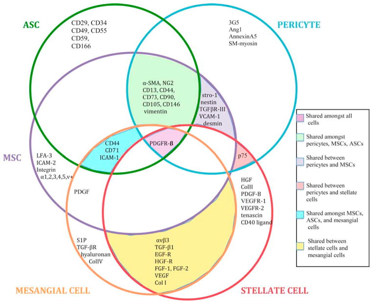
Pericytes are mural cells of the microcirculation that have been shown to play key roles in regulating microvascular morphogenesis and stability throughout each tissue bed and organ system assessed. Of note, recent work has revealed that pericytes share several characteristics with mesenchymal- and adipose-derived stem cells, suggesting there may be lineage-related connections among bona fide pericytes and these vascular "progenitors," which can assume a perivascular position in association with endothelial cells. Hence, pericyte identity as a mediator of vascular remodeling may be confounded by its close relationships with its progenitors or pluripotent cell counterparts and yet demonstrates their potential utility as cell-based therapies for unmet clinical needs. Crucial to the development of such therapies is a comprehensive understanding of the origin and fate regulating these related cell types as well as the unveiling of the molecular mechanisms by which pericytes and endothelial cells communicate. Such mechanistic inputs, which disrupt normal cellular crosstalk during disease inception and progression, offer opportunities for intervention and are discussed in the context of the vasculopathies accompanying tumor growth, diabetes, and fibrosis.
Copyright © 2014 Mosby, Inc. All rights reserved.
Conflict of interest statement
The authors have no financial or personal conflicts of interest that may influence the material presented in this review and recognize
Figures
References
-
- Armulik A, Genové G, Betsholtz C. Pericytes: Developmental, physiological, and pathological perspectives, problems, and promises. Dev Cell. 2011;21(2):193–215. - PubMed
-
- Sims DE. The pericyte – a review. Tissue Cell. 1986;18(2):153–174. - PubMed
-
- Armulik A, Abramsson A, Betsholtz C. Endothelial/pericyte interactions. Circ Res. 2005;97(6):512–23. - PubMed
-
- Díaz-Flores L, Gutiérrez R, Madrid JF, et al. Pericytes. Morphofunction, interactions and pathology in a quiescent and activated mesenchymal cell niche. Histol Histopathol. 2009;24(7):909–69. - PubMed
-
- von Tell D, Armulik A, Betsholtz C. Pericytes and vascular stability. Exp Cell Res. 2006;312(5):623–9. - PubMed
Publication types
MeSH terms
Grants and funding
LinkOut - more resources
Full Text Sources
Other Literature Sources
