. 2014 May 1:195:19-26.
doi: 10.1016/j.resp.2014.01.020. Epub 2014 Feb 13.

Effects of modulators of AMP-activated protein kinase on TASK-1/3 and intracellular Ca(2+) concentration in rat carotid body glomus cells

Affiliations

Effects of modulators of AMP-activated protein kinase on TASK-1/3 and intracellular Ca(2+) concentration in rat carotid body glomus cells

Donghee Kim et al. Respir Physiol Neurobiol. .

Abstract

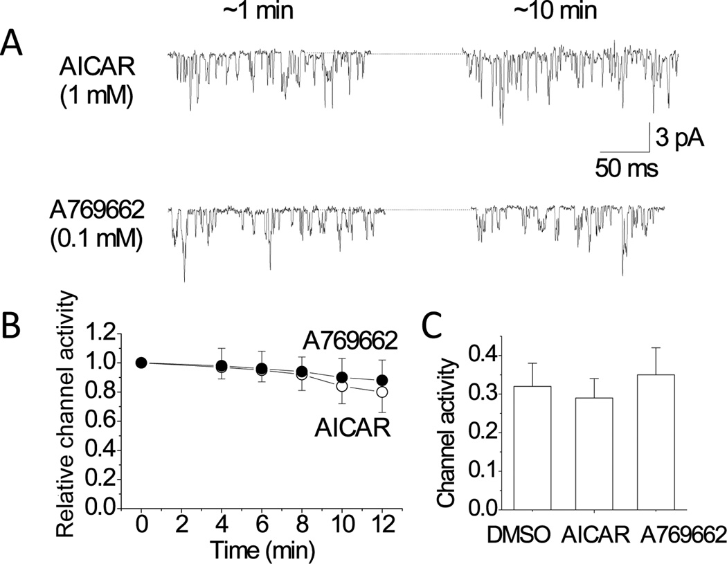
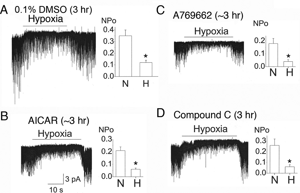
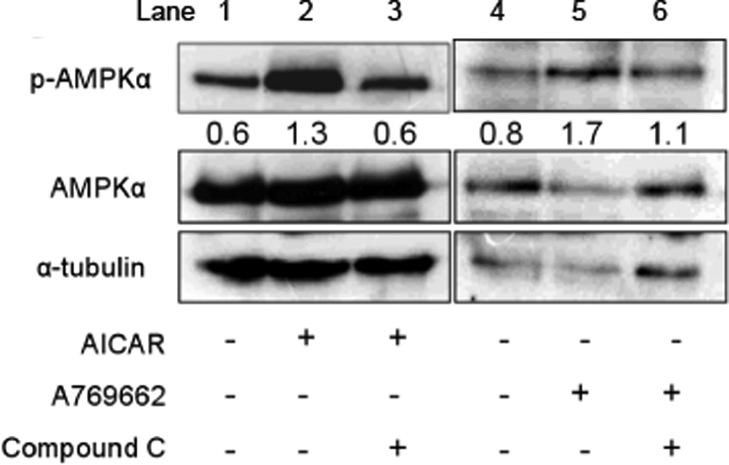
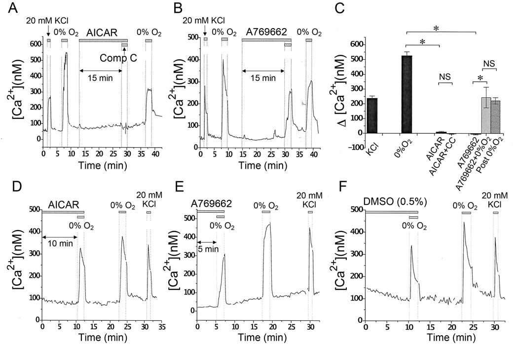
Acute hypoxia depolarizes carotid body chemoreceptor (glomus) cells and elevates intracellular Ca(2+) concentration ([Ca(2+)]i). Recent studies suggest that AMP-activated protein kinase (AMPK) mediates these effects of hypoxia by inhibiting the background K(+) channels such as TASK. Here we studied the effects of modulators of AMPK on TASK activity in cell-attached patches. Activators of AMPK (1mM AICAR and 0.1-0.5mM A769662) did not inhibit TASK activity or cause depolarization during acute (10min) or prolonged (2-3h) exposure. Hypoxia inhibited TASK activity by ∼70% in cells pretreated with AICAR or A769662. Both AICAR and A769662 (15-40min) failed to increase [Ca(2+)]i in glomus cells. Compound C (40μM), an inhibitor of AMPK, showed no effect on hypoxia-induced inhibition of TASK. AICAR and A769662 phosphorylated AMPKα in PC12 cells, and Compound C blocked the phosphorylation. Our results suggest that AMPK does not affect TASK activity and is not involved in hypoxia-induced elevation of intracellular [Ca(2+)] in isolated rat carotid body glomus cells.

Keywords: AMP kinase; Background K(+) channels; Carotid body; Chemoreceptors; Hypoxia.


Figures

Figure 1
Figure 1. No effect of AICAR and A769662 on TASK activity in glomus cells
A. Cell-attached patches were perfused with solution 1 mM AICAR or 0.1 mM A769662 until patches broke or up to 12 min. Pipette potential was set at 0 mV. Representative single-channel currents at ~1 min and ~10 min following application of the drugs are shown. B. TASK activity (NPo) was determined at various time points and plotted as a function of perfusion time. TASK activity was calculated and normalized to that obtained at time zero (1.0). Each point is the mean±SD of 12 determinations from 6 separate cell preparations. C. TASK activity in cell-attached patches was determined from cells incubated with 0.1% DMSO, 1 mM AICAR or 0.1 mM A769662 for 2–3 h. Each bar is the mean±SD of 12 patches.
Figure 2
Figure 2. AMPK modulators do not affect the hypoxia-induced inhibition of TASK activity
A. Cell-attached patches were perfused with normoxic solution, then with hypoxia solution for ~30 sec, and then back to normoxic solution. Glomus cells were incubated for 2–3 h with 0.1% DMSO. B-C. Same experiment as in A except that glomus cells were incubated with 1 mM AICAR (B) or A760662 (C) for 2–3 h before recording TASK in cell-attached patches. Perfusion solutions contained either AICAR or A769662. D. Glomus cells were incubated with 40 µM Compound C for 2–3 hr. Cell-attached patches were then formed and perfused with normoxic and hypoxic solution containing Compound C. For all bar graphs (A-D), each bar is the mean±SD of 6–8 patches. Asterisk (*) indicates a significant reduction from control (normoxia) at p < 0.05. For all experiments in this figure, data were gathered from 4 separate cell preparations.
Figure 3
Figure 3. Phosphorylation of AMPKα by AICAR and A769662
PC12 cells were treated with 1 mM AICAR or 100 µM A769662 for 60 min. Compound C (40 µM) was pretreated for 30 min to block AMPK activation. Activated AMPK (p-AMPKα) was detected in PC12 cells using a specific antibody for the phosphorylated form of AMPKα. Whole-cell lysate was used as total protein. Equal amounts (30 µg) of total protein were loaded in each lane. The plus and minus signs (+ and −) represent treatment conditions with and without chemical, respectively. The bands obtained from Western blot were quantified by Quantity One software (version 4.6.3) attached to GS-800 Calibrated densitometer (Bio-Rad). The numbers below the pAMPKα band represent the ratio of the expression levels of p-AMPKα to those of AMPKα for each lane. For example, in lanes 1and 4 (control), the level of phosphorylated p-AMPKα compared to that of AMPKα is 0.6 and 0.8, respectively. AICAR or A769662 increased the fractional levels of AMPKα phosphorylation (lanes 2 and 5) and Compound C blocked this effect (lanes 3 and 6). The α-tubulin was used as a loading control. The size of molecules: p-AMPKα (62 kDa), AMPKα (62 kDa), and α-tubulin (50 kDa). The experiment was done three times and an image from one experiment is shown.
Figure 4
Figure 4. AICAR and A769662 do not elevate intracellular [Ca2+]
A. Representative averaged trace of changes in [Ca2+]i in glomus cells in response to 20 mM KCl, hypoxia, AICAR (1 mM), and AICAR (1 mM) + Compound C (40 µM) are shown. B. Representative averaged trace of changes in [Ca2+]i in glomus cells in response to 20 mM KCl, hypoxia, A769662 (0.1 mM), and A769662 (0.1 mM) + 0% O2 are shown. C. Averaged [Ca2+]i increase above the basal level in response to 20 mM KCl (n=15), 0% O2 (n=15), 1 mM AICAR (n=15), AICAR (1 mM) + Compound C (40 µM) (n=15), A769662 (0.1 mM) (n=25), and A769662 (0.1 mM) + 0% O2 (n=8). Each bar represents the mean±SE. Asterisk(*) indicates significant difference at p < 0.001. NS= not significant. D-F. Glomus cells were incubated in medium containing 1 mM AICAR (D), 0.5 mM A769662 (E) or 0.5% DMSO (F) for 30 min at 37°C before recording [Ca2+]. Representative averaged traces of changes in [Ca2+]i in response to hypoxia and 20 mM KCl are shown. For these experiments, cells from 3 separate preparations were used.

References

    1. Bain J, Plater L, Elliott M, Shpiro N, Hastie CJ, McLauchlan H, Klevernic I, Arthur JS, Alessi DR, Cohen P. The selectivity of protein kinase inhibitors: a further update. Biochem J. 2007;408:297–315. - PMC - PubMed
    1. Buckler KJ. TASK-like potassium channels and oxygen sensing in the carotid body. Respir Physiol Neurobiol. 2007;157:55–64. - PubMed
    1. Buckler KJ. Effects of exogenous hydrogen sulphide on calcium signalling, background (TASK) K channel activity and mitochondrial function in chemoreceptor cells. Pflugers Arch. 2012;463:743–754. - PMC - PubMed
    1. Buckler KJ, Vaughan-Jones RD. Effects of mitochondrial uncouplers on intracellular calcium, pH and membrane potential in rat carotid body type I cells. The Journal of physiology. 1998;513((Pt 3)):819–833. - PMC - PubMed
    1. Corton JM, Gillespie JG, Hawley SA, Hardie DG. 5-aminoimidazole-4-carboxamide ribonucleoside. A specific method for activating AMP-activated protein kinase in intact cells? Eur J Biochem. 1995;229:558–565. - PubMed

Publication types

MeSH terms