. 2014 Mar;20(3):265-71.
doi: 10.1038/nm.3465. Epub 2014 Feb 16.

p38 MAPK signaling underlies a cell-autonomous loss of stem cell self-renewal in skeletal muscle of aged mice

Affiliations

p38 MAPK signaling underlies a cell-autonomous loss of stem cell self-renewal in skeletal muscle of aged mice

Jennifer D Bernet et al. Nat Med. 2014 Mar.

Abstract

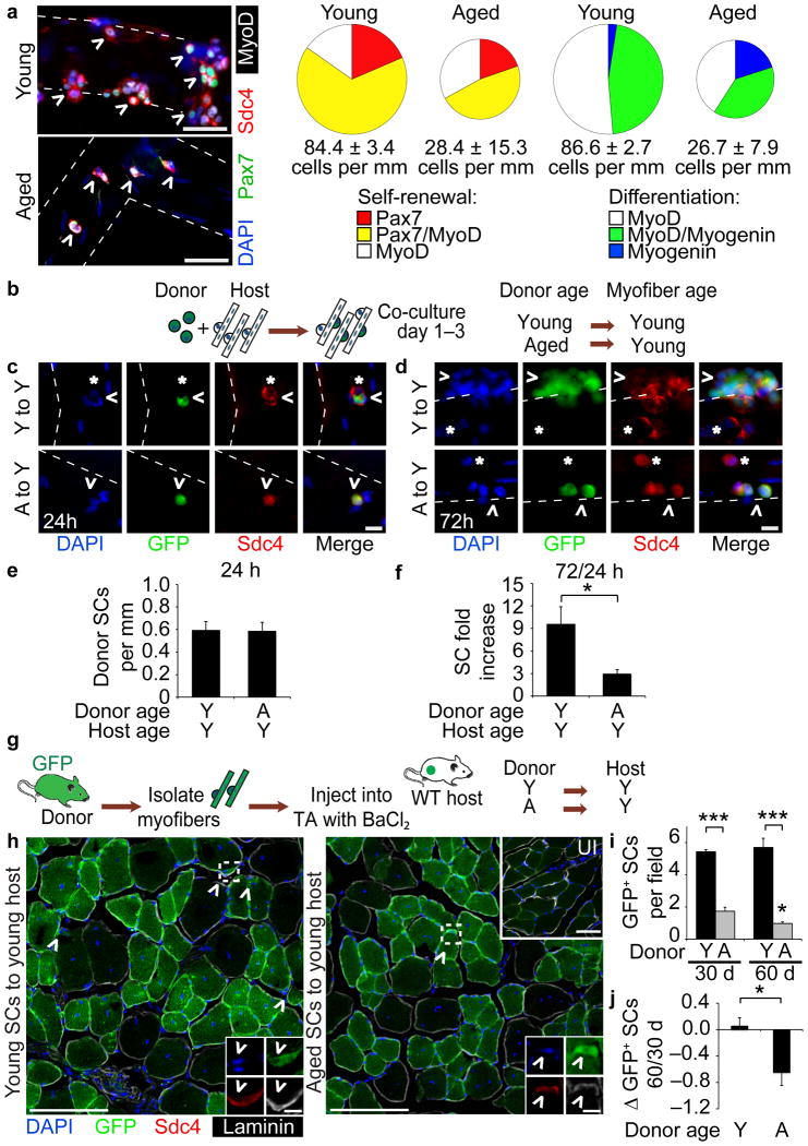
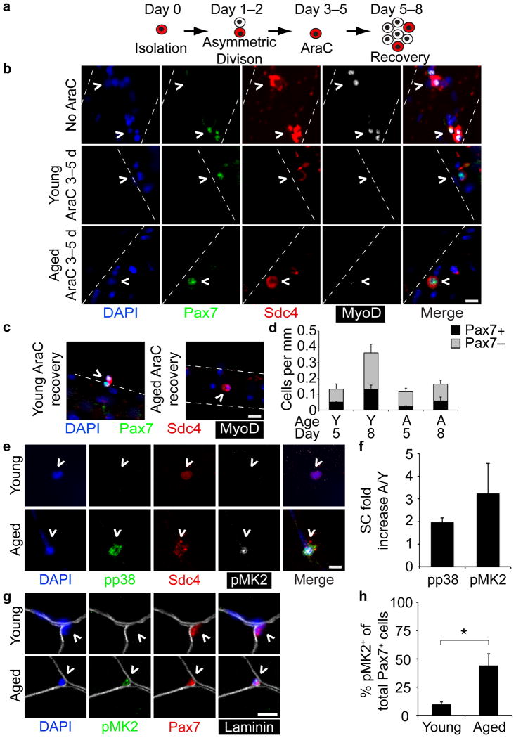
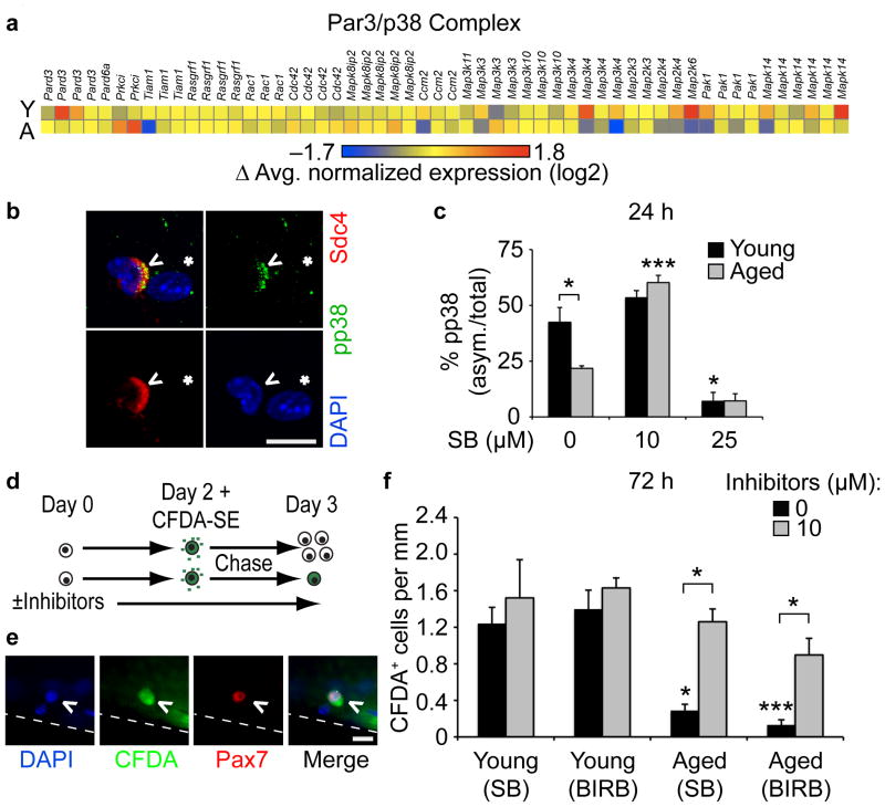
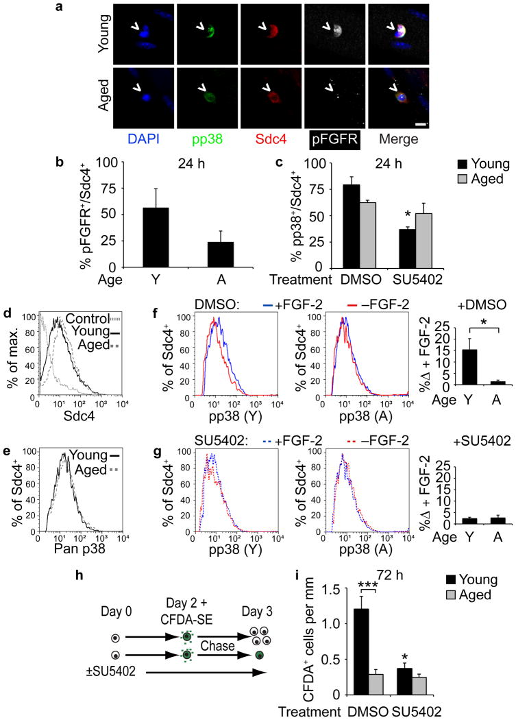
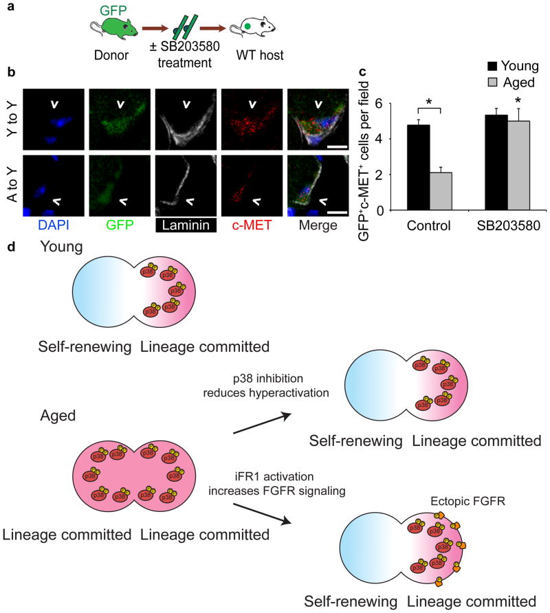
Skeletal muscle aging results in a gradual loss of skeletal muscle mass, skeletal muscle function and regenerative capacity, which can lead to sarcopenia and increased mortality. Although the mechanisms underlying sarcopenia remain unclear, the skeletal muscle stem cell, or satellite cell, is required for muscle regeneration. Therefore, identification of signaling pathways affecting satellite cell function during aging may provide insights into therapeutic targets for combating sarcopenia. Here, we show that a cell-autonomous loss in self-renewal occurs via alterations in fibroblast growth factor receptor-1, p38α and p38β mitogen-activated protein kinase signaling in satellite cells from aged mice. We further demonstrate that pharmacological manipulation of these pathways can ameliorate age-associated self-renewal defects. Thus, our data highlight an age-associated deregulation of a satellite cell homeostatic network and reveal potential therapeutic opportunities for the treatment of progressive muscle wasting.


Figures

Figure 1
Figure 1. Heterochronic transplantation of aged satellite cells (SCs) to local or systemic young environment fails to rescue age-associated phenotypes
(a) (left) Young and aged myofiber-associated SCs cultured for 96 h and stained with Pax7, MyoD and Myogenin. Scale bar, 50 μm. (right) Percentages of Syndecan-4+ (Sdc4+) SCs at 96 h that are quiescent (Pax7+), proliferating myoblasts (Pax7+/MyoD+ or MyoD+) or differentiating myocytes (MyoD+/Myogenin+ or Myogenin+). Chart area scaled to Sdc4+ SCs per myofiber length (n = 3, ≥ 20 myofibers/condition. P < 0.05 for 96 h Young vs. Aged cells/myofiber length and Pax7/MyoD+. P < 0.001 for MyoD/Myogenin+, t-test.). (b–f) (b) Schematic for heterochronic, βActGFP SC and wild-type myofiber co-culture. GFP+Sdc4+ SCs (^) and endogenous Sdc4+ SCs (*) after (c) 24 h or (d) 72 h in culture. Scale bar, 50 μm. Average number of GFP+/Sdc4+ SCs per myofiber length at (e) 24 h, and (f) fold increase in donor SCs per myofiber at 72 h in culture (n = 3, ≥ 20 myofibers per condition. *P < 0.05, t test). (g-j) (g) Schematic for heterochronic myofiber transplantation to young muscle hosts. (h) Muscle sections harvested at 30 d or 60 d (not shown) post-transplantation . Insets depict donor-derived SCs (^ = GFP+/Sdc4+) and contralateral, uninjured (UI) muscle. Scale bars, 50 μm or 5 μm (SCs). (i) Average number of donor-derived SCs per field (j) and Log2 fold-change in donor-derived SCs per field comparing 60 d vs. 30 d post-transplantation (n = 3 to 5 transplant recipients. *P < 0.05 for Aged 30 d vs. 60 d; ***P < 0.001 for Young vs. Aged, t test). Mean ± s.e.m. for all.
Figure 2
Figure 2. Loss of self-renewal in aged SCs correlates with elevated p38 signaling in aged SCs
(a–d) (a) Schematic for AraC treatment of myofibers to identify quiescent daughter SCs. (b) Young and aged myofibers treated for 48 h with or without AraC (Day 5) and (c) after additional 72 h recovery without AraC (Day 8). Scale bars,10 μm. (d) Average number of Pax7+, AraC-resistant SCs (^) and Pax7, terminally differentiated myocytes per myofiber length (P < 0.05 for Pax7+ Young 5 d vs. 8 d and total cells Young 5 d vs. 8 d, one-way ANOVA. ≥ 20 myofibers scored/condition). (e-h) young and aged (e) Sdc4+ myofiber-associated SCs (^) at 1 h post-dissection stained for phospho-p38 (pp38) and phospho-MK2 (pMK2) (aged SC is adjacent to myonucleus) and plotted (f) for the average fold increase in number of pp38+ and pMK2+ aged SCs/young SCs (≥20 myofibers scored/age). (g) Pax7+ SCs (^) in young and aged uninjured muscle sections stained for pMK2 and Pax7 (Scale bars, 10 μm) and plotted for (h) the percentage of activated SCs (Pax7+/pMK2+)/total SCs (Pax7+) (≥30 fields. *P < 0.05, t test). Mean ± s.e.m. and n = 3 for all.
Figure 3
Figure 3. Partial inhibition of p38αβ MAPK rescues aged SCs self-renewal
(a) Heatmap of normalized expression of proteins involved in asymmetric SC cell division. (b–c) (b) Myofiber-associated, asymmetric phospho-p38+ SC (^) near a myonucleus (*). Scale bar, 10 μm. (c) Percentage of asymmetric (asym.) phospho-p38+ SCs after 24 h culture treated with increasing SB203580 (SB) (*P < 0.05 for Young vs. Aged 0 μM SB, Young 0 vs. 25 μM SB; ***P < 0.001 for Aged 0 vs. 10 μM SB, (unmarked) Young 10 vs. 25 μM SB and Aged 10 vs. 25 μM SB, two-way ANOVA. Mean ± s.e.m., n = 3 experiments, ≥20 myofibers/condition). (d–f) (d) Schematic for CFDA-SE retention assay. (e) Self-renewed (Pax7+/CFDA-SE+), myofiber-associated SC. Scale bar, 10 μm. Scored and plotted (f) as average number of Pax7+/CFDA-SE+ SCs per myofiber length either untreated or treated with SB203580 or BIRB 796 (*P < 0.05 for Young vs. Aged 0 μM SB, Aged 0 vs. 10 μM (SB and BIRB); ***P < 0.001 for Young vs. Aged 0 μM BIRB, two-way ANOVA; mean ± s.e.m., n = 3 experiments, ≥30 myofibers).
Figure 4
Figure 4. FGFR1 signaling is altered in aged compared to young SCs
(a–c) (a) Young and aged Sdc4+ myofiber-associated SCs (^) cultured for 24 h and immunostained for pp38 and phospho-FGFR (pFGFR). Scale bar, 10 μm. (b) Percentage of pFGFR+ SCs out of total Sdc4+ SCs (Mean ± s.e.m., n = 3 experiments, ≥ 20 myofibers scored/condition). (c) Percentage of phospho-p38+ SCs of total Sdc4+ SCs after 24 h treatment with DMSO or SU5402 (*P < 0.05 for Young DMSO vs. SU5402, two-way ANOVA. n = 3 experiments, ≥ 20 myofibers scored/condition). (d–g) Flow cytometric analysis of young and aged pp38+ SCs with or without FGF-2 addition. Histograms of (d) percent of Sdc4+ events of total (max.) events and (e) p38αβ MAPK+ events of Sdc4+ subsets. Histograms of pp38+ SCs (gated on Sdc4+) following a 5 min FGF addition to (f) SCs in DMSO or (g) 25 μM SU5402 quantified (right) for FGF-2-induced increases in pp38+ (n = 3 experiments. *P < 0.05, t test). (h–i) (h) CFDA-SE retention assay identifies self-renewed (Pax7+/CFDA-SE+), myofiber-associated SCs. (i) Average number of Pax7+/CFDA-SE+ SCs per myofiber length after a 72 h treatment with DMSO or SU5402 (*P < 0.05 for Young DMSO vs. SU5402; ***P< 0.001 for Young vs. Aged DMSO, two-way ANOVA). Mean ± s.e.m. for all.
Figure 5
Figure 5. Constitutive FGFR1 signaling partially rescues self-renewal in aged SCs
(a–c) (a) Schematic for iFGFR1 (iFR1) transfection of young and aged SCs treated with or without Dimerizer B/B (B/B) to activate iFR1. (b) Image of iFR1+ SCs (^ = HA-tagged-iFR1). Scale bars, 50 μm; inset (box), 10 μm. (c) Percentage of Pax7+ or Myogenin+ (of total iFR1+ SCs) with or without Dimerizer B/B treatment (P < 0.05 for 0 vs.100 nM B/B (both Young and Aged), one-way ANOVA). (d-f) (d) Staining of pFGFR and (e) pp38 in young and aged iFR1+ SCs (^), treated with or without Dimerizer B/B for 48 h. Scale bar, 5 μm. (f) The percentage of asymmetric pp38, asymmetric iFR1 or co-localized, asymmetric pp38 and iFR1 Sdc4+ SCs (*P < 0.05, t test. Mean ± s.e.m., n = 3 experiments, ≥30 myofibers). (g-j) (g) Schematic for CFDA-SE retention assay of iFR1 expressing SCs. (h) Label-retaining, iFR1-transfected young and aged SCs (^ = CFDA+/Pax7+/iFR1+). Scale bar, 10 μm. (i) Average number of label-retaining SCs per myofiber length (*P < 0.05, for Young vs. Aged B/B treated, Young vs. Aged FGF-2 treated, Aged B/B treated vs. FGF-2 treated, two-way ANOVA) and (j) average fold-change for number of young and aged B/B treated label-retaining SCs vs. FGF-2 treated SCs (*P < 0.05, t test. Mean±s.e.m., n = 3 experiments, ≥20 myofibers).
Figure 6
Figure 6. Partial p38αβ MAPK inhibition rescues aged SC engraftment
(a–c) (a) Schematic for heterochronic myofiber transplantation into young host muscles after overnight treatment with 10 μM SB203580 or FGF-2 (control). (b) Images of donor-derived SCs (^ = GFP+/c-Met+) in muscle sections 30 d post-transplantation. Scale bar, 5 μm. (c) Average number of donor-derived SCs per field (Mean ± s.e.m. n = 3 transplant recipients. *P < 0.05 for Aged Control vs.SB treated, Young vs. Aged Control, two-way ANOVA). (d) Model. Asymmetric p38αβ MAPK activation promotes self-renewal in young SCs generating a quiescent daughter and a lineage committed daughter cell. Elevated pp38 in aged cells prevents asymmetric p38αβ MAPK signal transduction generating two lineage committed daughter cells. Partial inhibition of p38αβ MAPK permits asymmetric p38αβ MAPK activation, restoring self-renewal in aged SCs. Ectopic activation of constitutively expressed iFR1 re-localizes iFR1 into an asymmetric signaling complex promoting asymmetric localization of active iFR1, resulting in asymmetric activation of p38αβ MAPK and self-renewal.

Comment in

  • Rejuvenating aged muscle stem cells.
    Bentzinger CF, Rudnicki MA. Bentzinger CF, et al. Nat Med. 2014 Mar;20(3):234-5. doi: 10.1038/nm.3499. Nat Med. 2014. PMID: 24603790 No abstract available.

References

    1. Evans WJ, Campbell WW. Sarcopenia and age-related changes in body composition and functional capacity. 1993;123:465–468. - PubMed
    1. Baumgartner RN, et al. Epidemiology of sarcopenia among the elderly in New Mexico. 1998;147:755–763. - PubMed
    1. Roubenoff R. Sarcopenia: A major modifiable cause of frailty in the elderly: Sarcopenia in aging. 2000;4:140–142. - PubMed
    1. Landi F, et al. Sarcopenia and mortality risk in frail older persons aged 80 years and older: results from ilSIRENTE study. 2013;42:203–9. doi: 10.1093/ageing/afs194. - DOI - PubMed
    1. Janssen I, Shepard DS, Katzmarzyk PT, Roubenoff R. The healthcare costs of sarcopenia in the United States. 2004;52:80–5. - PubMed

Publication types

MeSH terms

Substances

Associated data