. 2014 May;63(5):437-48.
doi: 10.1007/s00262-014-1525-z. Epub 2014 Feb 15.

ImmTAC-redirected tumour cell killing induces and potentiates antigen cross-presentation by dendritic cells

Affiliations

ImmTAC-redirected tumour cell killing induces and potentiates antigen cross-presentation by dendritic cells

Giovanna Bossi et al. Cancer Immunol Immunother. 2014 May.

Abstract

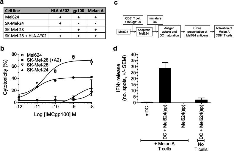
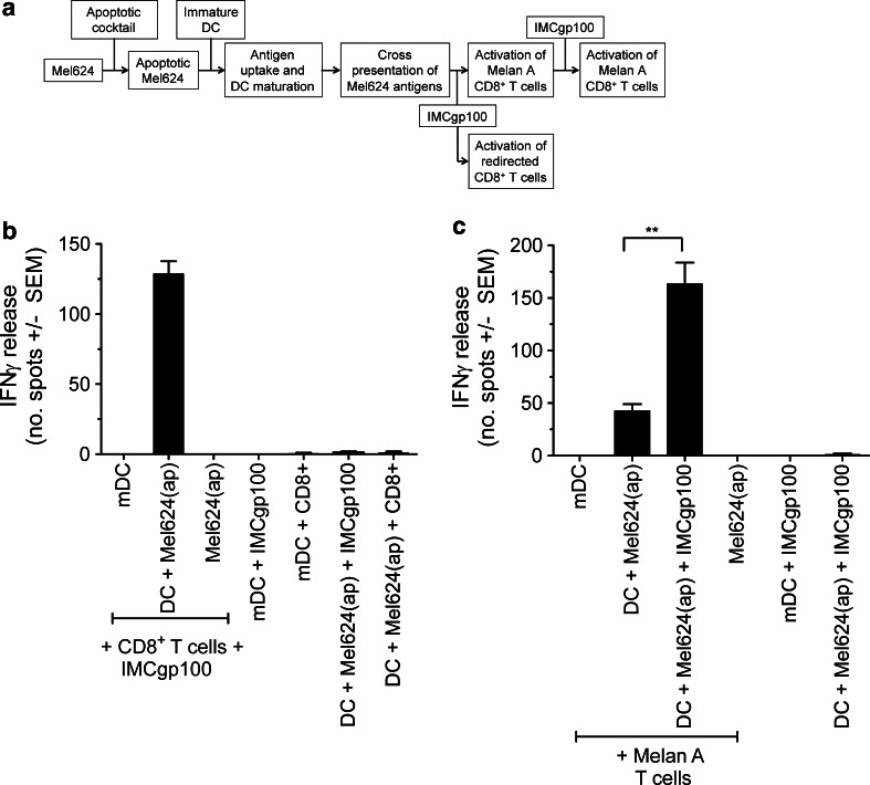
Antigen cross-presentation by dendritic cells (DCs) is thought to play a critical role in driving a polyclonal and durable T cell response against cancer. It follows, therefore, that the capacity of emerging immunotherapeutic agents to orchestrate tumour eradication may depend on their ability to induce antigen cross-presentation. ImmTACs [immune-mobilising monoclonal TCRs (T cell receptors) against cancer] are a new class of soluble bi-specific anti-cancer agents that combine pico-molar affinity TCR-based antigen recognition with T cell activation via a CD3-specific antibody fragment. ImmTACs specifically recognise human leucocyte antigen (HLA)-restricted tumour-associated antigens, presented by cancer cells, leading to T cell redirection and a potent anti-tumour response. Using an ImmTAC specific for a HLA-A*02-restricted peptide derived from the melanoma antigen gp100 (termed IMCgp100), we here observe that ImmTAC-driven melanoma-cell death leads to cross-presentation of melanoma antigens by DCs. These, in turn, can activate both melanoma-specific T cells and polyclonal T cells redirected by IMCgp100. Moreover, activation of melanoma-specific T cells by cross-presenting DCs is enhanced in the presence of IMCgp100; a feature that serves to increase the prospect of breaking tolerance in the tumour microenvironment. The mechanism of DC cross-presentation occurs via 'cross-dressing' which involves the rapid and direct capture by DCs of membrane fragments from dying tumour cells. DC cross-presentation of gp100-peptide-HLA complexes was visualised and quantified using a fluorescently labelled soluble TCR. These data demonstrate how ImmTACs engage with the innate and adaptive components of the immune system enhancing the prospect of mediating an effective and durable anti-tumour response in patients.


Conflict of interest statement

All authors are employees of Immunocore Ltd. Soluble monoclonal TCRs and ImmTACs, including IMCgp100, were produced by Immunocore Ltd.

Figures

Fig. 1
Fig. 1
IMCgp100-driven killing of melanoma cell lines leads to antigen cross-presentation by dendritic cells. a Details of the cell lines used in this study. b IMCgp100 dose–response curve showing redirected T cell cytotoxicity against the cell lines detailed in (a). Cytotoxicity was determined by LDH release after 24 h. Polyclonal CD8+ T cells, obtained from a healthy donor, were used at 100,000 cells per well at an effector target ratio of 10:1. Data shown represent the average of three independent measurements ± SEM. c Flow diagram summarising the experimental approach used to monitor IMCgp100-driven cross-presentation. d Apoptotic Mel624 cells [denoted Mel624(ap)] were produced by IMCgp100-redirected CD8+ T cell killing and subsequently incubated with immature DCs for 48 h to allow antigen uptake. IMCgp100 was used at a concentration of 0.1 nM. The resulting mature DCs were assessed for their ability to induce IFNγ release from Melan-A-specific CD8+ T cells. A further sample was prepared using LPS-matured DCs (mDC) only. Data shown represent the average of three independent measurements ± SEM. Additional control samples were prepared in a similar manner but in the absence of either DCs or Melan A T cells. These control measurements were performed in duplicate and are shown as average ± SEM
Fig. 2
Fig. 2
IMCgp100 redirects T cells in response to cross-presented antigen and enhances activation of TAA-specific T cells. a Flow diagram summarising the experimental approach used. b Apoptotic Mel624 cells [denoted Mel624(ap)] were prepared by chemically induced apoptosis and subsequently incubated with immature DCs for 48 h to allow antigen uptake. The resulting mature DCs were assessed for their ability to induce IFNγ release from IMCgp100-redirected autologous polyclonal CD8+ T cells. c The same approach was used to monitor activation of Melan-A-specific CD8+ T cells in the presence or absence of 0.1 nM IMCgp100. In each case, a further sample was prepared using LPS-matured DCs (mDC) only. Data shown represent the average of three independent measurements ± SEM. Additional control samples were prepared in a similar manner in the absence of various components as indicated on the graph. Control measurements were performed in duplicate and are shown as average ± SEM. Statistical significance was evaluated using an unpaired t test. p values were determined at the 95 % confidence interval
Fig. 3
Fig. 3
IMCgp100 elicits multiple effector functions. Experiments were performed using the approach shown in Fig. 2a. a and b DC-mediated activation of autologous polyclonal CD8+ (a) or CD4+ (b) T cell populations by IMCgp100 at concentrations between 10−13 and 10−8 M. A further sample prepared using LPS-matured DCs and ImmTAC-redirected T cells was measured concurrently. Data shown represent the average of three independent measurements ± SEM. Additional control experiments were carried out in the absence of IMCgp100, polyclonal T cells, DCs or apoptotic Mel624 cells, where necessary IMCgp100 was used at a fixed concentration of 1nM. c Poly-cytokine release from IMCgp100-redirected CD8+ T cells after incubation with DCs for 24 h. Intracellular accumulation of IFNγ, IL-2, TNF-α and MIP-1β, as well as the expression of lysosomal associated protein CD107a was detected by antibodies as described in the methods and analysed by Flow cytometry. Cells were first sorted by IFNγ production. A control measurement was made in the absence of IMCgp100
Fig. 4
Fig. 4
Cross-presented gp100-HLA-A*0201 complex is visualised and quantified on the DC cell surface. a A high-affinity biotinylated mTCR specific for the gp100-HLA-A*0201 complex was used to detect pHLA complexes on DCs after antigen uptake from chemically induced apoptotic Mel624 cells. An mTCR with alternative specificity was used as a control. Representative phase-contrast and fluorescence images are shown. The scale bar represents 20 μm. b The number of fluorescent spots on individual DCs was quantified manually from 2D Z-stack images. Additional control measurements were made using LPS-matured DCs, in the absence of apoptotic Mel624 cells. From left to right, the number of cells counted was 15, 54, 10 and 78. Statistical significance was evaluated using an unpaired t test. P values were determined at the 95 % confidence interval. c and d The same experiments analysis was performed using HLA-A*0201 DCs. From left to right, the number of cells counted was 9, 60, 6 and 22
Fig. 5
Fig. 5
T cell activation by DCs cross-presenting gp100 and Melan A is independent of the DC HLA restriction. DCs previously exposed to chemically induced apoptotic Mel624 cells [denoted Mel624(ap)] were assessed for their ability to induce IFNγ release from IMCgp100-redirected autologous polyclonal CD8+ T cells and Melan-A-specific CD8+ T cells (as described in Fig. 2a). IMCgp100 was used at a concentration of 1 nM. In this case, experiments were carried out using both HLA-A*0201+ (a) and HLA-A*0201 cross-presenting DCs (b). Further samples were prepared using LPS-matured DCs (mDC) only and in the absence of DCs. Data shown represent the average of three independent measurements ± SEM. The proliferation of IMCgp100-redirected CD8+ T cells was measured in response to HLA-A*0201+ (c) and HLA-A*0201 (d) cross-presenting DCs by CFSE labelling. The percentage of cells undergoing proliferation is indicated
Fig. 6
Fig. 6
Intact peptide-HLA complexes are acquired by DCs from apoptotic tumour cells. Immature HLA-A*0201+ DCs were exposed to chemically induced apoptotic melanoma cell lines, SK-Mel-28 (gp100+/Melan A+/HLA-A*0201), SK-Mel-24 (gp100/Melan A/HLA-A*0201+), Mel624 (gp100+/Melan A+/HLA-A*0201+) or SK-Mel-28 cells transduced with HLA-A*0201 (SK-Mel-28 A2+). The resulting DCs were assessed for their ability to induce IFNγ release from IMCgp100-redirected CD8+ polyclonal T cells and Melan-A-specific CD8 T cells. IMCgp100 was used at a concentration of 1 nM. A control measurement was made using LPS-matured DCs (mDC), in the absence of apoptotic Mel624 cells. Data shown represent the average of three independent measurements ± SEM

References

    1. Borghesi L, Milcarek C. Innate versus adaptive immunity: a paradigm past its prime? Cancer Res. 2007;67(9):3989–3993. doi: 10.1158/0008-5472.CAN-07-0182. - DOI - PubMed
    1. Ikeda H, Chamoto K, Tsuji T, Suzuki Y, Wakita D, Takeshima T, Nishimura T. The critical role of type-1 innate and acquired immunity in tumor immunotherapy. Cancer Sci. 2004;95(9):697–703. doi: 10.1111/j.1349-7006.2004.tb03248.x. - DOI - PMC - PubMed
    1. Celluzzi CM, Mayordomo JI, Storkus WJ, Lotze MT, Falo LD., Jr Peptide-pulsed dendritic cells induce antigen-specific CTL-mediated protective tumor immunity. J Exp Med. 1996;183(1):283–287. doi: 10.1084/jem.183.1.283. - DOI - PMC - PubMed
    1. Mackensen A, Meidenbauer N, Vogl S, Laumer M, Berger J, Andreesen R. Phase I study of adoptive T-cell therapy using antigen-specific CD8+ T cells for the treatment of patients with metastatic melanoma. J Clin Oncol. 2006;24(31):5060–5069. doi: 10.1200/JCO.2006.07.1100. - DOI - PubMed
    1. Spel L, Boelens J, Nierkens S, Boes M. Antitumor immune responses mediated by dendritic cells: how signals derived from dying cancer cells drive antigen cross-presentation. OncoImmunology. 2013;2:e26403. doi: 10.4161/onci.26403. - DOI - PMC - PubMed

Publication types

MeSH terms