Review
. 2014 May;137(Pt 5):1304-22.
doi: 10.1093/brain/awu002. Epub 2014 Feb 14.

Glucocerebrosidase is shaking up the synucleinopathies

Affiliations
Review

Glucocerebrosidase is shaking up the synucleinopathies

Marina Siebert et al. Brain. 2014 May.

Abstract

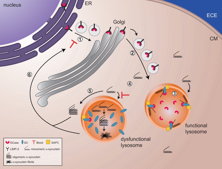
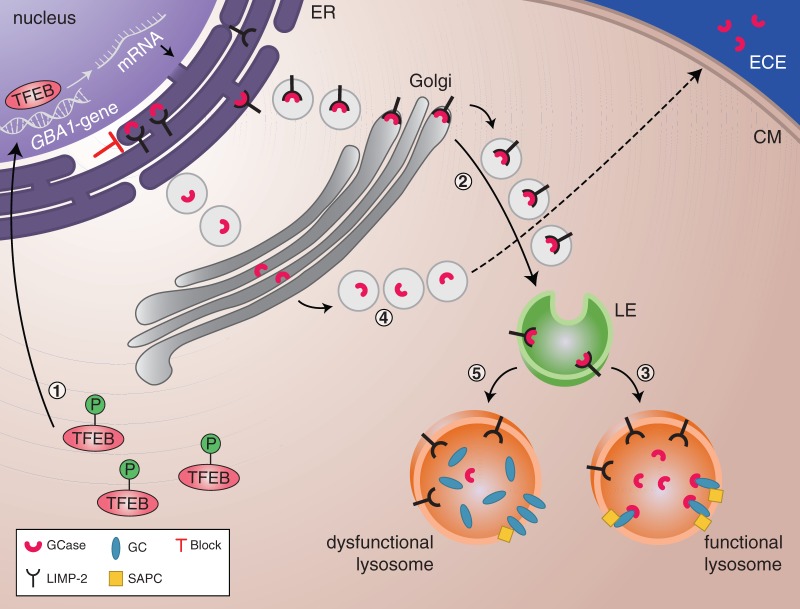
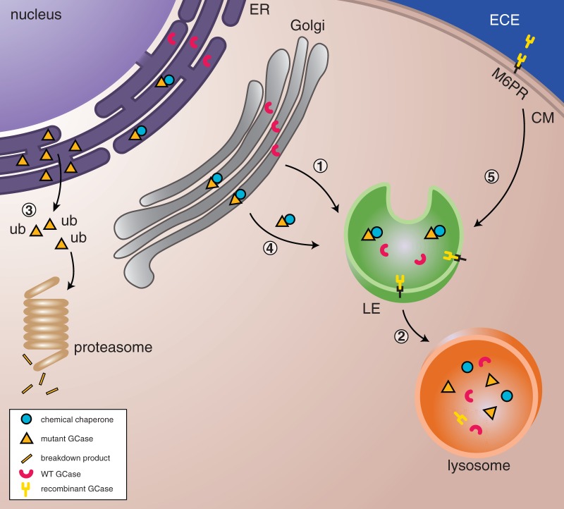
The lysosomal enzyme glucocerebrosidase, encoded by the glucocerebrosidase gene, is involved in the breakdown of glucocerebroside into glucose and ceramide. Lysosomal build-up of the substrate glucocerebroside occurs in cells of the reticulo-endothelial system in patients with Gaucher disease, a rare lysosomal storage disorder caused by the recessively inherited deficiency of glucocerebrosidase. Gaucher disease has a broad clinical phenotypic spectrum, divided into non-neuronopathic and neuronopathic forms. Like many monogenic diseases, the correlation between clinical manifestations and molecular genotype is not straightforward. There is now a well-established clinical association between mutations in the glucocerebrosidase gene and the development of more prevalent multifactorial disorders including Parkinson's disease and other synucleinopathies. In this review we discuss recent studies advancing our understanding of the cellular relationship between glucocerebrosidase and α-synuclein, the potential impact of established and emerging therapeutics for Gaucher disease for the treatment of the synucleinopathies, and the role of lysosomal pathways in the pathogenesis of these neurodegenerative disorders.

Keywords: Gaucher disease; Parkinson’s disease; glucocerebrosidase; lysosome; α-synuclein.


Figures

Figure 1
Figure 1
The reciprocal relationship between glucocerebrosidase and α-synuclein. (1 and 2) Glucocerebrosidase (GCase) is sorted via the endoplasmic reticulum and Golgi to the lysosomes. (3) In lysosomes, glucocerebrosidase interacts with its substrate glucocerebroside (GC) as well as monomers of α-synuclein, facilitating the breakdown of both at acidic pH. (4) Decreased levels of glucocerebrosidase will result in a slowdown of α-synuclein degradation and a gradual build up of glucocerebroside substrate, with the eventual formation of α-synuclein oligomers and fibrils. (4) Impaired lysosomes, as a result of build up of substrate and/or α-synuclein oligomers and fibrils, will show impaired chaperone-mediated autophagy and autophagosome fusion, which implies that α-synuclein cannot undergo autophagy-mediated degradation, resulting in an increased accumulation of α-synuclein in the cytoplasm. (5) These soluble monomers will eventually assemble in oligomers and will block trafficking of glucocerebrosidase from the endoplasmic reticulum (ER) to the Golgi. ECE = extracellular environment; CM = Cell Membrane; SAPC = saposin C.
Figure 2
Figure 2
Regulators of GBA1 expression and glucocerebrosidase activity and trafficking. (1) Phosphorylated TFEB is located in the cytoplasm. Under starvation conditions, dephosphorylated TFEB translocates to the nucleus where it regulates the transcription of genes involved in the CLEAR network, including GBA1. (2) GBA1 messenger RNA is translated into glucocerebrosidase. The interaction with its receptor LIMP-2 facilitates translocation of glucocerebrosidase to the lysosomes via the endoplasmic reticulum (ER), Golgi and late endosomes (LE). (3) In the lysosomes, glucocerebrosidase dissociates from LIMP-2. The glucocerebrosidase activator, saposin C (SAPC), lifts glucocerebroside from the lysosomal membrane and/or membranes within the lysosome and makes it available for glucocerebrosidase-mediated breakdown. (4) When LIMP-2 is deficient or absent, the glucocerebrosidase enzyme cannot be correctly sorted to the lysosomes. As a consequence, glucocerebrosidase is secreted into the extracellular environment (ECE). (5) Saposin C deficiency leads to the impairment of glucocerebroside degradation since the substrate is not available to glucocerebrosidase and subsequently accumulates inside the lysosomes. CM = Cell Membrane.
Figure 3
Figure 3
Therapeutic strategies to enhance glucocerebrosidase. (1 and 2) In healthy cells, wild-type glucocerebrosidase (WT GCase) is sorted to the lysosome via the endoplasmic reticulum, Golgi, and late endosomes (LE) where it will degrade its substrate glucocerebroside. (3) Mutant glucocerebrosidase is misfolded in the endoplasmic reticulum, becomes polyubiquitinated (ub) and undergoes proteasome-mediated degradation. (4) Pharmacological chaperones can stabilize mutant glucocerebrosidase and facilitate translocation to lysosomes. (5) In enzyme replacement therapy, recombinant glucocerebrosidase enzyme is delivered into the cells via the mannose-6-phosphate receptor and trafficked through the late endosomes to the lysosomes where it is able to degrade substrate. CM = Cell Membrane.

Comment in

References

    1. Abeliovich A, Schmitz Y, Farinas I, Choi-Lundberg D, Ho WH, Castillo PE, et al. Mice lacking alpha-synuclein display functional deficits in the nigrostriatal dopamine system. Neuron. 2000;25:239–52. - PubMed
    1. Alattia JR, Shaw JE, Yip CM, Prive GG. Direct visualization of saposin remodelling of lipid bilayers. J Mol Biol. 2006;362:943–53. - PubMed
    1. Alattia JR, Shaw JE, Yip CM, Prive GG. Molecular imaging of membrane interfaces reveals mode of beta-glucosidase activation by saposin C. Proc Natl Acad Sci USA. 2007;104:17394–9. - PMC - PubMed
    1. Alvarez-Erviti L, Rodriguez-Oroz MC, Cooper JM, Caballero C, Ferrer I, Obeso JA, et al. Chaperone-mediated autophagy markers in Parkinson disease brains. Arch Neurol. 2010;67:1464–72. - PubMed
    1. Alvarez-Erviti L, Seow Y, Schapira AH, Gardiner C, Sargent IL, Wood MJ, et al. Lysosomal dysfunction increases exosome-mediated alpha-synuclein release and transmission. Neurobiol Dis. 2011;42:360–7. - PMC - PubMed

Publication types