. 2014 Mar 27;507(7493):519-22.
doi: 10.1038/nature12978. Epub 2014 Feb 16.

Structure-based programming of lymph-node targeting in molecular vaccines

Affiliations

Structure-based programming of lymph-node targeting in molecular vaccines

Haipeng Liu et al. Nature. .

Abstract

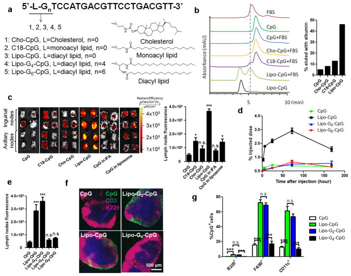
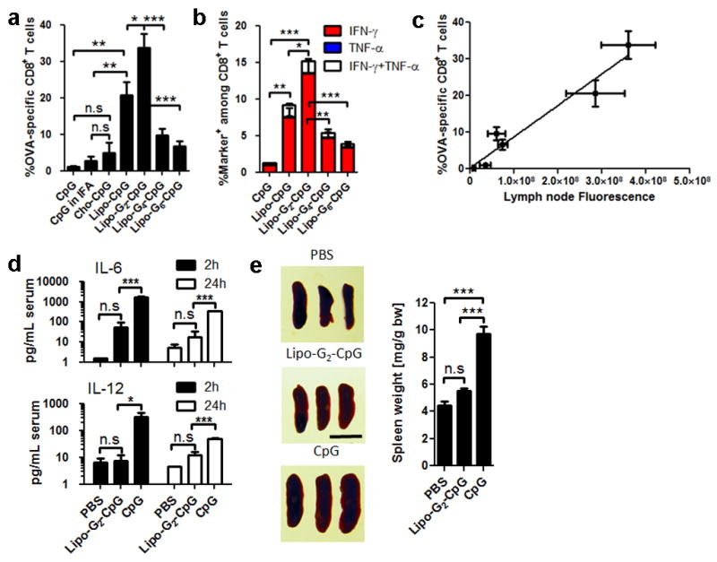
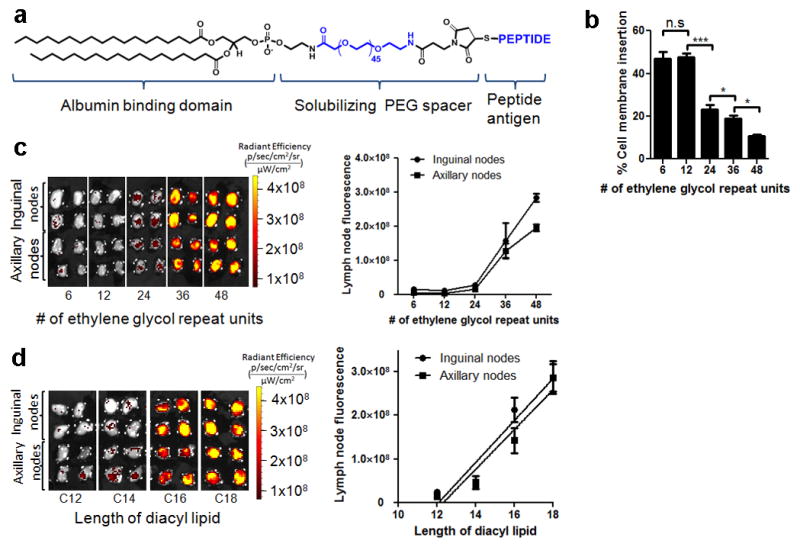
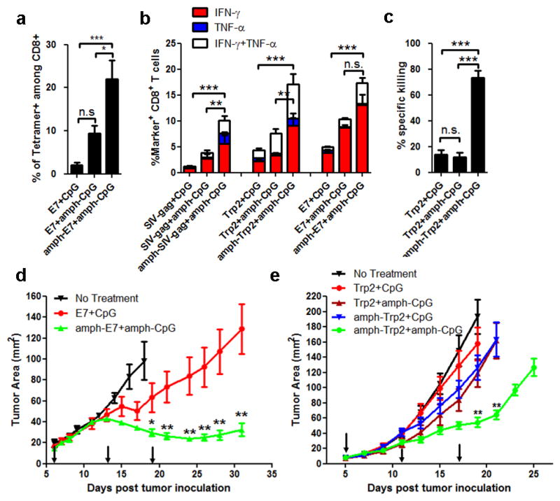
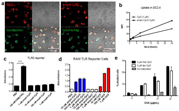
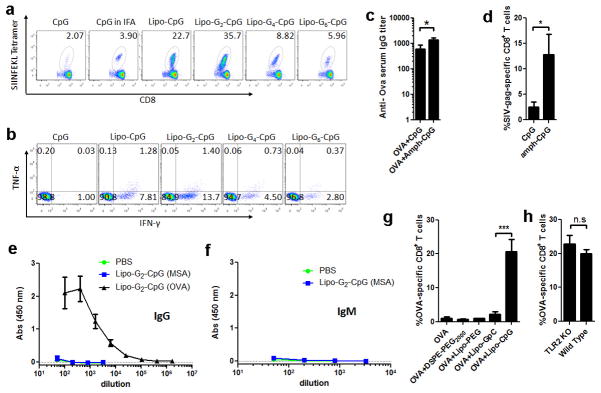
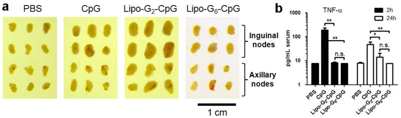
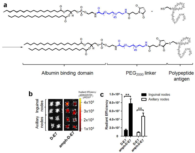
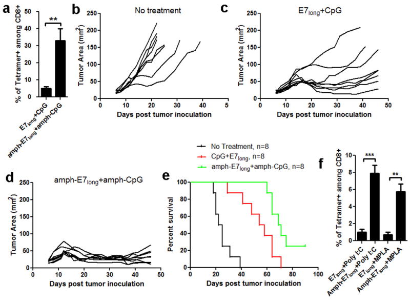
In cancer patients, visual identification of sentinel lymph nodes (LNs) is achieved by the injection of dyes that bind avidly to endogenous albumin, targeting these compounds to LNs, where they are efficiently filtered by resident phagocytes. Here we translate this 'albumin hitchhiking' approach to molecular vaccines, through the synthesis of amphiphiles (amph-vaccines) comprising an antigen or adjuvant cargo linked to a lipophilic albumin-binding tail by a solubility-promoting polar polymer chain. Administration of structurally optimized CpG-DNA/peptide amph-vaccines in mice resulted in marked increases in LN accumulation and decreased systemic dissemination relative to their parent compounds, leading to 30-fold increases in T-cell priming and enhanced anti-tumour efficacy while greatly reducing systemic toxicity. Amph-vaccines provide a simple, broadly applicable strategy to simultaneously increase the potency and safety of subunit vaccines.


Figures

Figure 1
Figure 1. Design of a lymph node-targeted molecular adjuvant
a, Structure of amph-CpGs. b, SEC of FAM-lCpGs alone or following incubation with FBS for 2h (left), and %CpG co-migrating with albumin peaks (right). Vertical dashed line provides a guide to the eye. c–g, IVIS fluorescence imaging of excised draining LNs from C57Bl/6 mice (n=4 LNs/group) injected with FAM-CpGs (3.3 nmol) in soluble form, emulsified in IFA, entrapped in liposomes, or as amphiphile conjugates. c, IVIS images and quantification from inguinal and axillary nodes at 24h. d, CpG accumulation in draining LNs. e, IVIS quantification of CpG in LNs 24h after injection of G-quadruplex-forming Lipo-Gn-CpGs. f, Immunohistochemistry of inguinal LNs 24h post-injection (CD3, blue; B220, pink; CpG, green). g, LN CpG+ cells determined by flow cytometry at 24h. ***, p 0.001; **, p<0.01; *, p<0.05 compared to soluble CpG by one-way ANOVA with Bonferroni post-test. Data represent mean±SEM of 2–3 independent experiments.
Figure 2
Figure 2. Lymph node targeting enhances the potency while reducing systemic toxicity of CpG
a–c, C57Bl/6 mice (n=4–8/group) were immunized with ovalbumin (10 μg) + CpG (1.24 nmol) on d0 and d14; shown are SIINFEKL tetramer (a) and intracellular cytokine staining (b) on peripheral blood at d20. c, LN CpG fluorescence correlation with T-cell response. d, Serum cytokines following injection (n=3/group) of 6.2 nmol CpG. e, Splenomegaly (n=3/group) assessed on d6 after 3 injections of CpG (scale bar: 1 cm). ***, p<0.001; **, p<0.01; *, p<0.05 by one-way ANOVA with Bonferroni post-test. Data show mean±SEM of 2–4 independent experiments.
Figure 3
Figure 3. Design of lymph node-targeted amphiphile-peptides
a, Structure of amph-peptides. b, Amph-PEG-fluorescein insertion into cell membranes quantified by fluorescence spectroscopy following 1h incubation with splenocytes in the presence of 100 μM albumin. c–d, C57Bl/6 mice (n=4 LNs/group) were injected with fluorescent amph-PEGs having varying PEG length (fixed C18 diacyl lipid tails, c) or lipid tail length (fixed PEG length 48 EG units, d); draining LNs were excised and imaged by IVIS after 24h. ***, p < 0.001; **, p < 0.01; *, p < 0.05 by one-way ANOVA with Bonferroni post-test. Data represent mean±SEM of two independent experiments.
Figure 4
Figure 4. Amph-vaccines maximize immunogenicity and therapeutic efficacy of polypeptide vaccines
a–c, C57Bl/6 mice (n=3–4/group) were immunized with SIV-gag, Trp2, or E7 peptides (10 μg) + CpG (1.24 nmol) on d0 and d14; shown are tetramer-positive CD8+ T-cells (a) and intracellular cytokine production (b) in peripheral blood on d20. c, Trp2-specific cytotoxicity measured using an in vivo killing assay on d21. d–e, Tumor growth in C57BL/6 mice (n=8/group) inoculated with 3×10⁵ TC-1 (d) or B16F10 (e) tumor cells and vaccinated with CpG + E7 peptide or Trp2 peptide (10 μg prime, 20 μg boost), respectively, on days indicated by arrows. Statistically-significant differences between soluble and amph-vaccines indicated by asterisks: ***, p<0.001; **, p<0.01; *, p<0.05 by one-way ANOVA with Bonferroni post-test. Shown are mean±SEM of 2–4 independent experiments.
Extended Data Figure 1
Extended Data Figure 1. Interaction between albumin and amph-CpGs
a, Size exclusion chromatography of fetal bovine serum (FBS), albumin, and fluorescein-labeled amph-CpGs. FBS and bovine serum albumin (BSA) were monitored using absorptions at 280 nm, while CpG oligos were monitored at 480 nm (Fluorescein peak). b, lipo-CpG, but not CpG, interact with serum albumin as shown by SDS-PAGE following protein pull-down assays: Fetal bovine serum (FBS) was incubated with 3′-biotin-labeled CpG (CpG-biotin), lipo-CpG (lipo-CpG-biotin), or PBS for 1 hr at 37°C. Streptavidin-conjugated magnetic beads were added to capture biotinylated CpGs and any associated proteins, separated by a magnet, boiled to release bound CpG/proteins, and subjected to SDS-PAGE analysis. Lane 1: protein MW ladder; lane 2: purified BSA; lane 3: FBS (100× dilution, 10 μL loading); lane 4: pull-down control, FBS incubated with streptavidin-magnetic beads; lane 5: pull-down with CpG-biotin, FBS was incubated with CpG-biotin and streptavidin-magetic beads; lane 6: pull-down with lipo-CpG-biotin, FBS incubated with lipo-CpG-biotin and streptavidin magnetic beads; lane 7: FBS (100× dilution, 5 μL loading). c, Fluorescein-labeled CpG or lipo-CpG was incubated with albumin-conjugated agarose resin for 1 hr at 37°C, and the resin was separated by filtration. The filtrate and recovered agarose were visualized by a gel imager and quantified by fluorescence measurements. d, Bio-layer interferometry measurements of lipo-CpG and CpG binding to immobilized bovine serum albumin. Albumin-conjugated BLI probes were immersed in solutions of lipo-CpG, and wavelength shifts (Δλ) of the interferometry pattern association and dissociation curves were followed over time to determine affinity constants of binding at 25°C. Shown are apparent kon, koff, and KD values from fits to the data. e, Fluorescence resonance energy transfer (FRET) between FAM-labeled lipo-CpG and rhodamine-conjugated albumin assessed by fluorescence spectroscopy. CpG-F or lipo-CpG-F (1.65 μM) alone or mixed with BSA-Rh (1.5 μM) in PBS were excited at 488 nm and emission was recorded from 500–650 nm.
Extended Data Figure 2
Extended Data Figure 2. Construction and characterization of G-quadruplex-stabilized CpG adjuvants
a, G-quadruplex stabilized CpG micelles are self-assembled from amphiphiles composed of three distinct segments: an immunostimulatory CpG sequence, a central repeat block containing n=1–10 G-quartet-forming guanines followed by (10–n) non-interacting thymidines, and a diacyl lipid tail. In aqueous solutions, these amphiphiles self-assembled into three-dimensional spherical micelles with a CpG corona and a lipid core. In the presence of K+, neighboring guanine repeats in the oligo corona form G-quadruplex structures via Hoogsteen hydrogen bonds and stabilize the micelle structure. The oligo micelles’ stabilities in the presence of serum were programmed by altering the length of the guanine repeat. b, Parallel G-quartet formation among DNA strands within the micelles was detected by circular dichroism (CD) spectroscopy, as manifested by the shifting of positive peaks from 278 nm toward 262 nm and troughs at 245 nm as the number of guanines in the structure increased. c, Pyrene excimer fluorescence was used to assay the stabilities of G-quadruplex micelles in the presence of albumin: Pyrene dye incorporated in stabilized CpG micelles (n > 2) retained excimer fluorescence in the presence of high concentrations of albumin. In contrast, albumin binds to the lipids moiety of unstabilized micelles (n ≤ 2) and disrupts the micelle structures, leading to loss of excimer fluorescence in an albumin concentration-dependent manner as the protein disrupts the micelles into albumin-bound unimers. Shown below the schematic is the fraction of amphiphile-CpG remaining in the micellar state as a function of albumin concentration as reported by excimer fluorescence. Arrow indicates the plasma concentration of albumin. d, Stability profiles of G-quadruplex CpG micelles as measured by size-exclusion chromatography in the presence of fetal bovine serum (FBS). Fluorescein-labeled CpG micelles were incubated with 20% FBS in PBS in the presence of 10 mM Mg2+ and 20 mM K+ at 37°C for 2 hours, then analyzed by SEC. FBS and BSA were monitored using absorptions at 280 nm, while lipo-Gn-CpG amphiphiles were monitored at 480 nm (Fluorescein peak). Lipo-Gn-CpG with n=0 or 2 partitioned to co-migrate with albumin, while amphiphiles with n>2 showed increasing fractions of the amphiphiles migrating as intact micelles in the presence of serum with increasing n. e, Fluorescein-labeled lipo-G6-CpG (5 μM) and Alexa Fluor® 647-labeled BSA (5 μM) were incubated for 2 hrs at 37 °C in PBS+20 mM KCl, and then analyzed by size exclusion chromatography. Spectra were monitored at 480 nm (ODN channel, green line, fluorescein) and 640 nm (protein channel, red line, Alexa Fluor® 647). The majority of BSA and CpG micellar aggregates eluted separately. f, Amph-CpG micelles sizes as determined by dynamic light scattering. All data are mean±s.e.m. Statistical analysis was performed by one-way ANOVA with Bonferroni post-tests.
Extended Data Figure 3
Extended Data Figure 3. Lymph node localization of amph-CpGs with macrophages and dendritic cells
a, Immunofluorescent images of inguinal LN section 24 h after injection of 3.3 nmol lipo-CpG or lipo-G2-CpG, showing dendritic cells (CD11c, blue), macrophages (F4/80, red), and CpG (green). b–d, Mice (n=3/group) were injected s.c. with 3.3 nmol of Fluorescein-labeled CpG formulations. After 24 h, lymph nodes were digested and lymph nodes cells stained with DAPI and antibodies against F4/80, CD11c and CD207. Shown are representative flow cytometry plots of F4/80 staining (b) and CD11c staining (c) versus CpG fluorescence in viable (DAPI) cells. d, percentages of CpG+ cells in the LNs determined by flow cytometry at 24 hr. ***, p < 0.001; All data are mean±s.e.m. Statistical analysis was performed by unpaired student’s t-test.
Extended Data Figure 4
Extended Data Figure 4. CpG-albumin conjugates accumulate in LNs
C57Bl/6 mice (n=4 LNs/group) were injected s.c. at the tail base with 3.3 nmol fluorescein-labeled free CpG, mouse albumin-CpG conjugates (MSA-CpG), or lipo-CpG. Inguinal LNs and axillary LNs were isolated 24 hours post injection and imaged (a) and quantified (b) by IVIS optical imaging. All data are mean±s.e.m. **, p < 0.01 by one-way ANOVA with Bonferroni post-test.
Extended Data Figure 5
Extended Data Figure 5. In-vitro characterization of amph-CpG
a, Rhodamine labeled CpG or amph-CpG (1 μM) was incubated with murine bone marrow-derived dendritic cells (BMDCs) at 37°C for 4 hours with LysoTracker® (Life Technologies) and imaged using a Zeiss LSM 510 confocal microscope (Oberkochen, Germany). b, Rhodamine labeled CpG or amph-CpG (1 μM) was incubated for 30 minutes, 2 hours, 6 hours, and 24 hours with the murine dendritic cell line DC2.4. Cells were stained with DAPI and uptake was quantified by flow cytometry using the mean fluorescence intensity (MFI) of viable (DAPI-) cells. c, Amph-CpG or PAM2CSK4 (a strong TLR2 agonist) was incubated for 24 hours with the InvivoGen HEK-Blue murine TLR2 reporter cell line, a secreted embryonic alkaline phosphatase (SEAP) reporter system. SEAP levels were quantified by incubating supernatant with QuantiBlue substrate for 1h and reading absorption at 620 nm. d, Amph-CpG, CpG, or control amph-GpC (1 μM) were incubated with InvivoGen RAW-Blue mouse macrophage reporter cells, which secrete SEAP upon TLR, NOD or Dectin-1 stimulation. SEAP levels were quantified by incubating supernatant with QuantiBlue substrate for 1h and reading absorption at 620 nm. e, Bone marrow-derived immature dendritic cells were incubated overnight with indicated concentrations of OVA and maturation stimuli (or medium alone). DCs were washed 3 times with PBS and 30,000 CFSE-labeled OT-I CD8+ T cells were then added to each well. Cells were collected after 2 days of co-culture, and stained and gated for DAPI- (viable) CD8+ T cells using Flowjo v.7.6.5 (Treestar). Extent of proliferation was quantified by determining the % of cells that had undergone division by determining % of viable CD8+ T cells that had diluted CFSE using T cells alone as a control for the no division/dilution peak. Shown are mean±s.e.m. in b–d; ***, p < 0.001 by one-way ANOVA with Bonferroni post-test. Bars in e represent medians and whiskers represent range (n=2 wells/condition).
Extended Data Figure 6
Extended Data Figure 6. Albumin-binding lipo-CpGs elicit robust expansion of antigen-specific CD8+ T-cells when combined with soluble protein
Groups of C57Bl/6 mice (n=4–8/group) were immunized s.c. on day 0 and day 14 with 10 μg OVA and 1.24 nmol CpG formulations as indicated. Six days after the final immunization, mice were bled and PBMCs were evaluated by SIINFEKL-tetramer staining and intracellular cytokine staining. a, Representative flow cytometric dot plots of H-2Kb/SIINFEKL tetramer staining of CD8+ cells. b, Representative flow dot plots of intracellular staining on CD8+ cells for IFN-γ and TNF-α after 6 h ex-vivo restimulation with SIINFEKL peptide. c, serum samples were collected and assayed by ELISA for anti-OVA IgG (day 34). d, mice were immunized on day 0 and day 14 with 1.24 nmol lipo-G2-CpG mixed with 10 μg SIV-gag protein, blood samples were collected and analyzed by peptide-MHC tetramer staining for CD8+ T-cells recognizing the immunodominant AL11 epitope of gag. e, f, anti-mouse serum albumin (MSA) and anti-OVA IgG (e, day 20) or IgM (f, day 20) were measured by ELISA. g, groups of C57Bl/6 mice (n=4/group) were immunized with 10 μg OVA alone or mixed with 1.24 nmol of a non-TLR agonist lipo-GpC, the same diacyl lipid tail conjugated to PEG (lipo-PEG, 48 EG units), or DSPE-PEG2000. Animals were boosted with the same formulation on day 14, and OVA tetramer+ CD8+ T-cells in peripheral blood were assayed by flow cytometry on day 20. ***, p < 0.001. h, TLR2 knockout or wild type mice were immunized as described in (a), and OVA tetramer+ CD8+ T-cells were assayed as previously. All data are mean±s.e.m. *, p < 0.05. Statistical analysis was performed by unpaired student’s t-test.
Extended Data Figure 7
Extended Data Figure 7. Albumin-binding CpG induces local lymphadenopathy but reduces systemic toxicity compared to soluble CpG adjuvant
a, C57Bl/6 mice (n=3/group) were injected with 1.24 nmol CpGs subcutaneously on day 0 and 2.48 nmol CpGs on days 2 and 4. On day 6 animals were sacrificed and lymph nodes were isolated and photographed with a digital camera. b, Bead-based flow analysis of proinflammatory cytokines elicited in peripheral blood of mice injected with a single dose (6.2 nmol) of different CpG formulations, blood samples were collect at different time interval and analyzed for TNF-α per manufacturer’s instructions. All data are mean±s.e.m. **, p < 0.01; *, p < 0.05. Statistical analysis was performed by one-way ANOVA with Bonferroni post-test.
Extended Data Figure 8
Extended Data Figure 8. Hydrophilic block length of amphiphiles determines cell membrane insertion and lymph node accumulation
a, Amphiphiles with varying hydrophilic PEG lengths were prepared by solid phase synthesis of diacyl tails coupled to 1–8 hexa-ethylene glycol phosphorothioate units. Fluorescein was incorporated either at the 3′ terminal (for membrane insertion analyses) or adjacent to the lipid moiety (for albumin-binding analyses). b, Splenocytes from C57Bl/6 mice (5×10⁷cells/mL) were incubated with lipo-(PEG)n-Fluorescein (1.67 μM) and albumin (100 μM) at 37°C for 1 hour. Shown is a representative image of membrane insertion observed by confocal microcopy for lipo-(PEG)1-fluorescein. c, Equilibrium partitioning measurements shown as a function of albumin concentration at 37°C. Lipo-fluorescein-(PEG)n (5 μM) was incubated with varying concentrations of BSA and fluorescence intensities were monitored by fluorescence spectroscopy. BSA binding disrupted the micellar structure and decreased the self-quenching of fluorescein. All samples reached maximum fluorescence intensities at around 10 μM BSA, indicating 100% micelle breakup at this concentration (at higher BSA concentrations, fluorescence decreases due to solution turbidity). Arrow indicates plasma concentration of albumin. d, e, Lipo-Tn-FAM amphiphiles (n=5, 10, 15, 20) were injected s.c. in C57Bl/6 mice (n=4 LNs/group) and excised LNs were imaged after 24 hr (d). Mean LN fluorescence from groups of mice are plotted in (e). All data are mean±s.e.m.
Extended Data Figure 9
Extended Data Figure 9. Lipo-PEG-peptide amphiphiles exhibit greatly enhanced lymph node accumulation compared to unmodified peptides
a, Peptides with N-terminal cysteines were conjugated to maleimide-PEG2000-DSPE.b–c, FAM-labeled immunodominant peptide derived from the HPV-16 E7 protein (FAM-FTVINYHARC, synthesized in reverse sequence order using D-amino acids to obtain the same chiral organization of side chains as the typical L-amino acid sequence in a protease-resistant peptide) was injected s.c. at the tail base as a free peptide (D-E7) or as a PEG-DSPE conjugate (amph-D-E7). Shown are IVIS images of draining lymph nodes 24 hours post injection (b) and fluorescence quantifications (c). All data are mean±s.e.m. **, p < 0.01. Statistical analysis was performed by unpaired student’s t-test.
Extended Data Figure 10
Extended Data Figure 10. Long peptide amphiphiles, when combined with amph-CpG, elicit potent antigen specific CD8+ T cells response with therapeutic benefits, as compared to soluble formulation
a, C57Bl/6 mice were primed on day 0 and boosted on day 14 with amph-E7long (HPV-16 E743–62, 10 μg peptide) and amph-CpG (Lipo-G2-CpG, 1.24 nmol) or equivalent soluble peptide/CpG vaccines. Six days post boost, mice were bled and analyzed for tetramer positive CD8+ T-cells in peripheral blood. b–e, C57BL/6 mice (n=8/group) were inoculated with 3×10⁵ TC-1 tumor cells s.c. in the flank and left untreated or immunized with soluble or amphiphile long peptide vaccines on days 6 (10 μg peptide, 1.24 nmol CpG), 13 (20 μg peptide, 1.24 nmol CpG), and 19 (20 μg peptide, 1.24 nmol CpG). Shown are individual tumor growth curves for no treatment (b), immunization with soluble E7long and CpG (c), or immunization with amph-E7long+amph-CpG (d). Kaplan-Meier survival curves of eight mice per group are shown in (e). f, long peptide amphiphiles also elicit potent immune responses when combined with non-CpG, non-lymph node targeting alternative adjuvants. C57Bl/6 mice (n=4/group) were immunized as before, using amph-E7long peptide (10 μg) combined with monophosphoryl lipid A (MPLA, 10 μg) or Polyinosinic:polycytidylic acid (Poly I:C, 50 μg). The frequencies of E7 tetramer+ CD8+ T-cells in peripheral blood were assayed on day 20. All data are mean±s.e.m. ***, p < 0.001; **, p < 0.01; *, p < 0.05 by unpaired student’s t-test.

References

    1. Salhab M, Patani N, Mokbel K. Sentinel lymph node micrometastasis in human breast cancer: an update. Surg Oncol. 2011;20:E195–E206. - PubMed
    1. Tsopelas C, Sutton R. Why certain dyes are useful for localizing the sentinel lymph node. J Nucl Med. 2002;43:1377–1382. - PubMed
    1. Johansen P, Mohanan D, Martínez-Gómez JM, Kündig TM, Gander B. Lympho-geographical concepts in vaccine delivery. J Controlled Release. 2010;148:56–62. - PubMed
    1. Moon JJ, Huang B, Irvine DJ. Engineering nano- and microparticles to tune immunity. Adv Mater. 2012;24:3724–3746. - PMC - PubMed
    1. Gupta RK. Aluminum compounds as vaccine adjuvants. Adv Drug Deliv Rev. 1998;32:155–172. - PubMed

Publication types