. 2014 Apr;42(8):5314-21.
doi: 10.1093/nar/gku128. Epub 2014 Feb 14.

TcaR-ssDNA complex crystal structure reveals new DNA binding mechanism of the MarR family proteins

Affiliations

TcaR-ssDNA complex crystal structure reveals new DNA binding mechanism of the MarR family proteins

Yu-Ming Chang et al. Nucleic Acids Res. 2014 Apr.

Abstract

The teicoplanin-associated locus regulator (TcaR) regulates gene expression of proteins on the intercellular adhesion (ica) locus involved in staphylococci poly-N-acetylglucosamine biosynthesis. The absence of TcaR increases poly-N-acetylglucosamine production and promotes biofilm formation. Until recently, the mechanism of multiple antibiotic resistance regulator family protein members, such as TcaR, was restricted to binding double-stranded DNA. However, we recently found that TcaR strongly interacts with single-stranded DNA, which is a new role for this family of proteins. In this study, we report Staphylococcus epidermidis TcaR-single-stranded DNA complex structures. Our model suggests that TcaR and single-stranded DNA form a 61-symmetry polymer composed of TcaR dimers with single-stranded DNA that wraps outside the polymer and 12 nt per TcaR dimer. Single-stranded DNA binding to TcaR involves a large conformational change at the DNA binding lobe. Several point mutations involving the single-stranded DNA binding surface validate interactions between single-stranded DNA and TcaR. Our results extend the novel role of multiple antibiotic resistance regulator family proteins in staphylococci.


Figures

Figure 1.
Figure 1.
The overall TcaR–ssDNA complex structure. (A) The crystal includes three TcaR dimers and one monomer per asymmetric unit. The TcaR dimers A/B (colored in green/cyan) and C/D (colored in yellow/magenta) interact with ssDNA, whereas the dimer E/F (colored in gray/orange) does not. (B) The final 2Fo-Fc omit density map with the nucleic acids contoured at a 1δ level is shown in gray mesh. The β-wing region is labeled with red ovals.
Figure 2.
Figure 2.
Structural comparison of the TcaR–ssDNA complex. (A) (Left) The TcaR apo (red) and ssDNA complex (green) structures are superimposed in cartoon mode. The TcaR complexes show significant conformational changes at the wHTH domain. (Right) The TcaR complexes are shown after an ∼90° rotation along the horizontal axis relative to the plane of the paper. (B) (Left) The apo TcaR dimer surfaces are colored red for Chain A and brown for Chain B. (Right) The TcaR–ssDNA complex surfaces are colored green for Chain A and blue for Chain B.
Figure 3.
Figure 3.
(A) The TcaR-24-nt-ssDNA complex model. The model was constructed based on the crystal structure of the complex formed by the TcaR dimers A/B (colored in green/cyan) and C/D (color in yellow/magenta). (B) Structural comparison of the dimer–dimer interactions in the TcaR apo (red) and ssDNA complex structures.
Figure 4.
Figure 4.
Model of the TcaR-long-chain–ssDNA complex. (A) Detailed interactions between TcaR dimers A/B (colored in green/cyan) and C/D (colored in yellow/magenta). (B and C) Stereo view of the combined model for the ssDNA location in the TcaR filament, as shown along the helical c axis in the space group P61 (b: bottom view; c: side view). The counterclockwise extended TcaR filament is right-handed with six TcaR molecules per turn and a 0.76 -Å rise per nucleotide. The TcaR filament sequence is red-green-blue-cyan-magenta-yellow. (D) Proposed model of the TcaR-long-chain–ssDNA complex. The complex has a 54.68 -Å helical pitch with a 132 -Å diameter. Each TcaR dimer interacts with 12 nt.
Figure 5.
Figure 5.
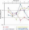
RT-PCR analysis of TcaR transcripts in S. epidermidis RP62A under different growth conditions. Relative levels of TcaR expressions were calculated compared with those of the control, 16S rRNA. The mean ± SD for the quantitative RT-PCR data is shown (n = 3).

References

    1. Anderson SD, Gums JG. Ceftobiprole: an extended-spectrum anti-methicillin-resistant Staphylococcus aureus cephalosporin. Ann. Pharmacother. 2008;42:806–816. - PubMed
    1. Brandenberger M, Tschierske M, Giachino P, Wada A, Berger-Bachi B. Inactivation of a novel three-cistronic operon tcaR-tcaA-tcaB increases teicoplanin resistance in Staphylococcus aureus. Biochim. Biophys. Acta. 2000;1523:135–139. - PubMed
    1. Jefferson KK, Pier DB, Goldmann DA, Pier GB. The teicoplanin-associated locus regulator (TcaR) and the intercellular adhesin locus regulator (IcaR) are transcriptional inhibitors of the ica locus in Staphylococcus aureus. J. Bacteriol. 2004;186:2449–2456. - PMC - PubMed
    1. McCallum N, Bischoff M, Maki H, Wada A, Berger-Bachi B. TcaR: a putative MarR-like regulator of sarS expression. J. Bacteriol. 2004;186:2966–2972. - PMC - PubMed
    1. Tegmark K, Karlsson A, Arvidson S. Identification and characterization of SarH1: a new global regulator of virulence gene expression in Staphylococcus aureus. Mol. Microbiol. 2000;37:398–409. - PubMed

Publication types

MeSH terms

Associated data