. 2014 Feb 14;343(6172):764-8.
doi: 10.1126/science.1244392.

Evolutionarily dynamic alternative splicing of GPR56 regulates regional cerebral cortical patterning

Affiliations

Evolutionarily dynamic alternative splicing of GPR56 regulates regional cerebral cortical patterning

Byoung-Il Bae et al. Science. .

Abstract

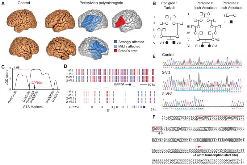
The human neocortex has numerous specialized functional areas whose formation is poorly understood. Here, we describe a 15-base pair deletion mutation in a regulatory element of GPR56 that selectively disrupts human cortex surrounding the Sylvian fissure bilaterally including "Broca's area," the primary language area, by disrupting regional GPR56 expression and blocking RFX transcription factor binding. GPR56 encodes a heterotrimeric guanine nucleotide-binding protein (G protein)-coupled receptor required for normal cortical development and is expressed in cortical progenitor cells. GPR56 expression levels regulate progenitor proliferation. GPR56 splice forms are highly variable between mice and humans, and the regulatory element of gyrencephalic mammals directs restricted lateral cortical expression. Our data reveal a mechanism by which control of GPR56 expression pattern by multiple alternative promoters can influence stem cell proliferation, gyral patterning, and, potentially, neocortex evolution.


Figures

Fig. 1
Fig. 1. A noncoding mutation in the GPR56 gene disrupts perisylvian gyri
(A) MRI shows polymicrogyria in the perisylvian area, in which abnormally thin cortex is folded in on itself, giving a paradoxical, but characteristic, thickened appearance (8). (B) Pedigrees of the three families with perisylvian polymicrogyria. (C) Linkage analysis isolates an interval containing GPR56. LOD, logarithm of the odds ratio for linkage. (D) The mutation arose independently in the Turkish and the Irish-American families. Haplotype mapping shows that pedigree 1 (1-V:1, 1-V:2) and pedigree 2 (2-VI:1, 2-VI:2) are unrelated. Homozygous single-nucleotide polymorphisms (SNPs) are shown in red or blue, heterozygous SNPs in green, and SNPs for which no genotype could be assigned in gray. (E and F) A homozygous deletion in one of two 15-bp tandem repeats (blue underscore and red box) upstream of GPR56 e1m causes perisylvian polymicrogyria. 2-V:2 stands for a heterozygous parent and 2-VI:2 for an affected individual from pedigree 2.
Fig. 2
Fig. 2. The noncoding mutation ablates lateral gene expression
(A) Schematic of the human GPR56 locus showing 17 alternative transcription start sites. E1m is highly expressed in the human fetal brain [mRNA-sequencing (mRNA-Seq) track, arrow]. The 15-bp deletion is upstream of e1m, located within a cis-regulatory element as one of two tandem 15-bp repeats. (B) A 23-kb upstream region of human GPR56 drives GFP expression throughout the transgenic mouse neocortex (E14.5), which mirrors endogenous GPR56 protein expression. The 15-bp deletion eliminates GFP expression from lateral cortex but preserves medial cortex expression, consistent with lesions observed by brain MRI (fig. S1) (n = 4 to 6 embryos with identical patterns per construct). Scale bar, 200 μm. (C) Y1H screening reveals Rfx transcription factor binding to the cis-regulatory element. See text for details. (D) The mutation decreases RFX binding to the cis-regulatory element in vitro. (E) RFX1 and GPR56 are colocalized in a human fetal brain 19 weeks after conception. Higher magnification of the outer subventricular zone is shown (right). v, ventricular zone; is, inner subventricular zone; os, outer subventricular zone; and i, intermediate zone. Scale bars, 100 μm (left) and 10 μm (right). (F) Dominant-negative RFX (white bars) abrogates normal e1m promoter activity. Black bars, GFP control. (G) Each RFX gene has distinct expression patterns in the fetal human brain. Each number means the corresponding RFX isoform. RFX3 and RFX7 are enriched in regions affected by perisylvian polymicrogyria (green boxes). pfc, prefrontal cortex; opfc, orbital pfc; dlpfc, dorsolateral pfc, mpfc, medial pfc; vlpfc, ventrolateral pfc; ms, motor-sensory cortex. *P < 0.001, t test.
Fig. 3
Fig. 3. GPR56 regulates neuroprogenitor proliferation
(A) In Gpr56 knockout mice, neurons overmigrate through breached pial basement membrane (arrowheads) or undermigrate (arrows) forming irregular cortical layers, as shown by immunostaining of Cux1, an upper layer (II to IV) marker (p9). Thin cortex is occasionally observed (asterisks). (B) GPR56 is highly expressed in human ventricular zone and outer subventricular zone at 12 to 17 weeks of gestation (GW), which suggests roles in neuroprogenitors. v, ventricular zone; s, subventricular zone; is, inner subventricular zone; os, outer subventricular zone; i, intermediate zone; c, cortical plate; and m, marginal zone. (C to D) Human GPR56 transgenic (Tg) mice have significantly more mitotic (PH3+) neuroprogenitor cells and intermediate progenitor (TBR2+) cells than wild-type (WT). In contrast, Gpr56 knockout (KO) mice have significantly fewer mitotic cells and intermediate progenitors than WT (E13.5 to E14.5). (n = 7 mice per genotype; *P < 0.005; **P < 0.001; paired t test). (E) The cells that are in utero electroporated (from E13.5 to E15.5) with human GPR56-IRES-GFP [either side of the internal ribosome entry site (IRES), GFP expressing] persist in the germinal zones longer that the GFP control cells. Red, TBR2; blue, Hoechst. (n = 11 mouse embryos per construct; *P < 0.0001; chi-squared test). Scale bars, 500 μm (A) and 100 μm (B) to (E).
Fig. 4
Fig. 4. GPR56 gene evolution
(A) The mouse Gpr56 locus has only 5 transcription start sites compared with 17 in human. (B) A 300-bp human GPR56 e1m promoter sequence containing the cis-regulatory element (Fig. 2A) directs β-gal expression to lateral cortex in mice (arrow), whereas the orthologous mouse e1c(m) promoter directs more widespread expression. Orthologous promoters from other mammals with larger cortical sizes, abundant gyri, or both (marmoset, dolphin, and cat) drive relatively limited expression patterns generally similar to human (n = 3 to 10 embryos with identical patterns per promoter). Scale bar, 2 mm. (C) Human, marmoset, dolphin, and cat brains are gyrencephalic or near-gyrencephalic with a Sylvian fissure (arrowhead). Mouse brain lacks both gyri and Sylvian fissure. Scale bar, 1 cm. Images from the University of Wisconsin and Michigan State Comparative Mammalian Brain Collections and/or the National Museum of Health and Medicine are reproduced with permission from Brainmuseum.org.

Comment in

References

    1. Rakic P. Nat Rev Neurosci. 2009;10:724–735. - PMC - PubMed
    1. Lui JH, Hansen DV, Kriegstein AR. Cell. 2011;146:18–36. - PMC - PubMed
    1. Zilles K, Palomero-Gallagher N, Amunts K. Trends Neurosci. 2013;36:275–284. - PubMed
    1. Amunts K, et al. PLOS Biol. 2010;8:e1000489. - PMC - PubMed
    1. Golden JA, Harding BN. Nat Rev Neurol. 2010;6:471–472. - PubMed

Publication types

MeSH terms