

New skin test for detection of bovine tuberculosis on the basis of antigen-displaying polyester inclusions produced by recombinant Escherichia coli
- PMID: 24532066
- PMCID: PMC3993176
- DOI: 10.1128/AEM.04168-13
New skin test for detection of bovine tuberculosis on the basis of antigen-displaying polyester inclusions produced by recombinant Escherichia coli
Abstract
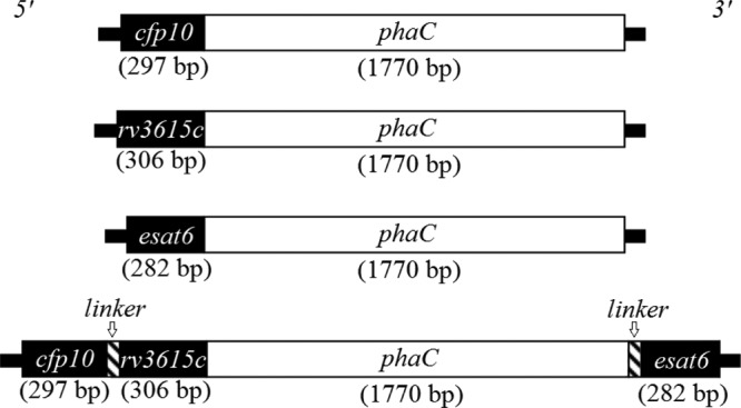
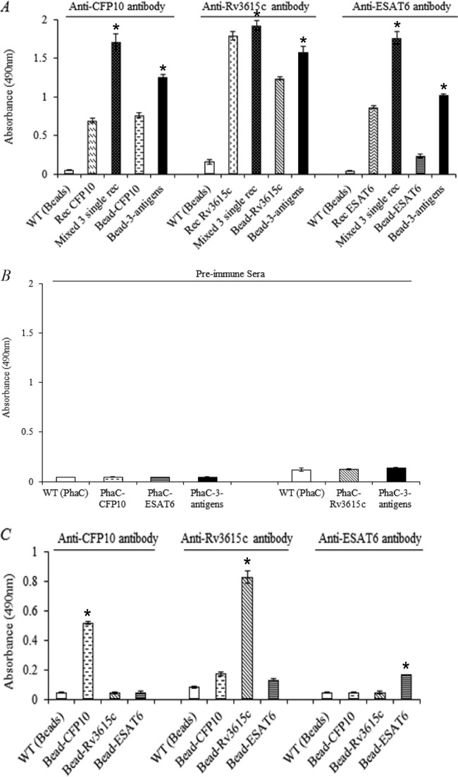
The tuberculin skin test for diagnosing tuberculosis (TB) in cattle lacks specificity if animals are sensitized to environmental mycobacteria, as some antigens in purified protein derivative (PPD) prepared from Mycobacterium bovis are present in nonpathogenic mycobacteria. Three immunodominant TB antigens, ESAT6, CFP10, and Rv3615c, are present in members of the pathogenic Mycobacterium tuberculosis complex but absent from the majority of environmental mycobacteria. These TB antigens have the potential to enhance skin test specificity. To increase their immunogenicity, these antigens were displayed on polyester beads by translationally fusing them to a polyhydroxyalkanoate (PHA) synthase which mediated formation of antigen-displaying inclusions in recombinant Escherichia coli. The most common form of these inclusions is poly(3-hydroxybutyric acid) (PHB). The respective fusion proteins displayed on these PHB inclusions (beads) were identified using tryptic peptide fingerprinting analysis in combination with matrix-assisted laser desorption ionization-time of flight mass spectrometry (MALDI-TOF MS). The surface exposure and accessibility of antigens were assessed by enzyme-linked immunosorbent assay (ELISA). Polyester beads displaying all three TB antigens showed greater reactivity with TB antigen-specific antibody than did beads displaying only one TB antigen. This was neither due to cross-reactivity of antibodies with the other two antigens nor due to differences in protein expression levels between beads displaying single or three TB antigens. The triple-antigen-displaying polyester beads were used for skin testing of cattle and detected all cattle experimentally infected with M. bovis with no false-positive reactions observed in those sensitized to environmental mycobacteria. The results suggested applicability of TB antigen-displaying polyester inclusions as diagnostic reagents for distinguishing TB-infected from noninfected animals.
Figures
Similar articles
-
Engineering Mycobacteria for the Production of Self-Assembling Biopolyesters Displaying Mycobacterial Antigens for Use as a Tuberculosis Vaccine.Appl Environ Microbiol. 2017 Feb 15;83(5):e02289-16. doi: 10.1128/AEM.02289-16. Print 2017 Mar 1. Appl Environ Microbiol. 2017. PMID: 28087528 Free PMC article.
-
Display of Antigens on Polyester Inclusions Lowers the Antigen Concentration Required for a Bovine Tuberculosis Skin Test.Clin Vaccine Immunol. 2015 Oct 28;23(1):19-26. doi: 10.1128/CVI.00462-15. Print 2016 Jan. Clin Vaccine Immunol. 2015. PMID: 26512049 Free PMC article.
-
Assessment of a protein cocktail-based skin test for bovine tuberculosis in a double-blind field test in cattle.Clin Vaccine Immunol. 2013 Apr;20(4):482-90. doi: 10.1128/CVI.00592-12. Epub 2013 Jan 30. Clin Vaccine Immunol. 2013. PMID: 23365203 Free PMC article.
-
Novel particulate vaccines utilizing polyester nanoparticles (bio-beads) for protection against Mycobacterium bovis infection - a review.Vet Immunol Immunopathol. 2014 Mar 15;158(1-2):8-13. doi: 10.1016/j.vetimm.2013.04.002. Epub 2013 Apr 30. Vet Immunol Immunopathol. 2014. PMID: 23707076 Review.
-
[Evolution of IGRA researches].Kekkaku. 2008 Sep;83(9):641-52. Kekkaku. 2008. PMID: 18979999 Review. Japanese.
Cited by
-
Mycobacterium avium subsp. paratuberculosis antigens induce cellular immune responses in cattle without causing reactivity to tuberculin in the tuberculosis skin test.Front Immunol. 2023 Jan 18;13:1087015. doi: 10.3389/fimmu.2022.1087015. eCollection 2022. Front Immunol. 2023. PMID: 36741398 Free PMC article.
-
A systematic review on the distribution of Mycobacterium bovis infection among wildlife in the Americas.Trop Anim Health Prod. 2019 Sep;51(7):1801-1805. doi: 10.1007/s11250-019-01954-7. Epub 2019 Jun 13. Trop Anim Health Prod. 2019. PMID: 31197725
-
Proteomic analysis of protein purified derivative of Mycobacterium bovis.J Transl Med. 2017 Apr 3;15(1):68. doi: 10.1186/s12967-017-1172-1. J Transl Med. 2017. PMID: 28372590 Free PMC article.
-
Engineering Mycobacteria for the Production of Self-Assembling Biopolyesters Displaying Mycobacterial Antigens for Use as a Tuberculosis Vaccine.Appl Environ Microbiol. 2017 Feb 15;83(5):e02289-16. doi: 10.1128/AEM.02289-16. Print 2017 Mar 1. Appl Environ Microbiol. 2017. PMID: 28087528 Free PMC article.
-
Bioengineering a bacterial pathogen to assemble its own particulate vaccine capable of inducing cellular immunity.Sci Rep. 2017 Feb 2;7:41607. doi: 10.1038/srep41607. Sci Rep. 2017. PMID: 28150705 Free PMC article.
References
-
- Cousins DV. 2001. Mycobacterium bovis infection and control in domestic livestock. Rev. Sci. Tech. 20:71–85 - PubMed
-
- Casal C, Bezos J, Diez-Guerrier A, Alvarez J, Romero B, de Juan L, Rodriguez-Campos S, Vordermeier M, Whelan A, Hewinson RG, Mateos A, Dominguez L, Aranaz A. 2012. Evaluation of two cocktails containing ESAT-6, CFP-10 and Rv-3615c in the intradermal test and the interferon-gamma assay for diagnosis of bovine tuberculosis. Prev. Vet. Med. 105:149–154. 10.1016/j.prevetmed.2012.02.007 - DOI - PubMed
Publication types
MeSH terms
Substances
LinkOut - more resources
Full Text Sources
Other Literature Sources
Medical
