. 2014 Sep;37(9):1707-15.
doi: 10.1007/s00449-014-1143-3. Epub 2014 Feb 16.

Liquid perfluorochemical-supported hybrid cell culture system for proliferation of chondrocytes on fibrous polylactide scaffolds

Affiliations

Liquid perfluorochemical-supported hybrid cell culture system for proliferation of chondrocytes on fibrous polylactide scaffolds

Maciej Pilarek et al. Bioprocess Biosyst Eng. 2014 Sep.

Abstract

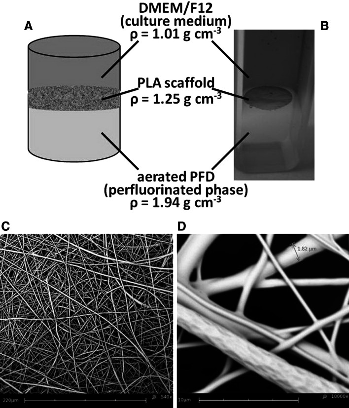
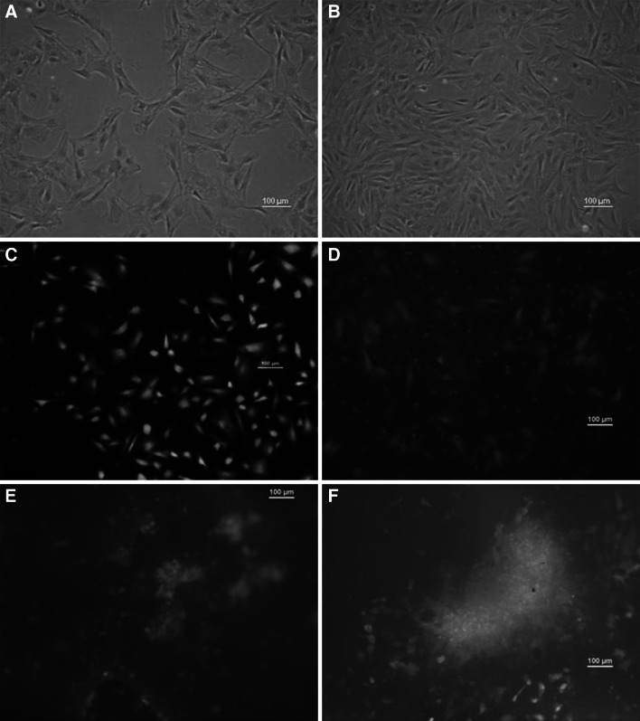
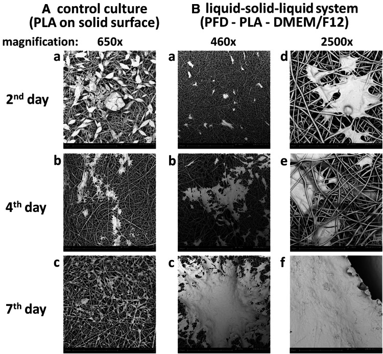
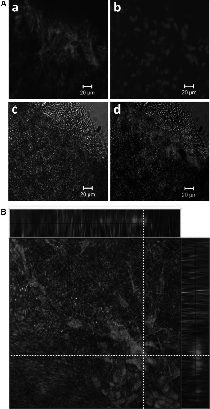
CP5 bovine chondrocytes were cultured on biodegradable electrospun fibrous polylactide (PLA) scaffolds placed on a flexible interface formed between two immiscible liquid phases: (1) hydrophobic perfluorochemical (PFC) and (2) aqueous culture medium, as a new way of cartilage implant development. Robust and intensive growth of CP5 cells was achieved in our hybrid liquid-solid-liquid culture system consisting of the fibrous PLA scaffolds in contrast to limited growth of the CP5 cells in traditional culture system with PLA scaffold placed on solid surface. The multicellular aggregates of CP5 cells covered the surface of PLA scaffolds and the chondrocytes migrated through and overgrew internal fibers of the scaffolds. Our hybrid culture system simultaneously allows the adhesion of adherent CP5 cells to fibers of PLA scaffolds as well as, due to use of phase of PFC, enhances the mass transfer in the case of supplying/removing of respiratory gases, i.e., O2 and CO2. Our flexible (independent of vessel shape) system is simple, ready-to-use and may utilize a variety of polymer-based scaffolds traditionally proposed for implant development.


Figures

Fig. 1
Fig. 1
The hybrid liquid–solid–liquid culture system containing liquid phase of air-saturated PFD, solid phase of fibrous PLA scaffold and liquid phase of DMEM:F12 medium: the outline (a), the example of hybrid culture system (b), SEM microphotography of PLA scaffold (c; scale bar 220 μm) and magnified fibrous PLA scaffold with marked diameter of single electrospun PLA fiber (d; scale bar 10 μm)
Fig. 2
Fig. 2
The comparison of the cells morphology of CP5 cells cultured in monolayer: 1st (a) and 7th (b) day of culture; cells cultured on the PLA scaffold placed directly on the bottom of the well (i.e. the control culture): 1st (c) and 7th (d) day of culture; cells cultured in the hybrid liquid–solid–liquid culture system: 2nd (e) and 7th (f) day of culture. a, b cells without staining; c, d, e, f live/dead fluorescence stained cells (green and orange colors for viable and dead cells, respectively). All scale bars 100 μm (color figure online)
Fig. 3
Fig. 3
SEM micrographs documenting the morphology of CP5 cells cultured on the PLA fibrous scaffold placed directly on the solid polystyrene surface of culture plate, i.e. the control culture A: 2nd (a), 4th (b) and 7th (c) day of culture; and cells cultured in the PFD supported hybrid liquid–solid–liquid culture system (B): 2nd (a, d), 4th (b, e) and 7th (c, f) day of culture. Scale bar represents: Aa Ab Ac 180 μm, Ba Bb Bc 250 μm, Bd Be Bf 40 μm
Fig. 4
Fig. 4
CLSM micrographs of CP5 chondrocytes growing inside the fibrous PLA scaffold taken from the liquid–solid–liquid culture system (7th day of culture): CP5 cells stained for actin molecules (Aa) and the cell nucleus DNA (Ab), the PLA fibers visible in transmitted light (Ac) and image of CP5 cells on the PLA scaffold (Ad); overview of the focal plane inside the PLA scaffold (B) (analyzed focal plane has been marked as white-dotted lines). All scale bars 20 μm (color figure online)

References

    1. Fassnacht D, Portner R. Experimental and theoretical considerations on oxygen supply for animal cell growth in fixed-bed reactors. J Biotechnol. 1999;72:169–184. doi: 10.1016/S0168-1656(99)00129-7. - DOI - PubMed
    1. Kellner K, Liebsch G, Klimant I, Wolfbeis OS, Blunk T, Schulz MB, Gopferich A. Determination of oxygen gradients in engineered tissue using a fluorescent sensor. Biotechnol Bioeng. 2002;80:73–83. doi: 10.1002/bit.10352. - DOI - PubMed
    1. Harrison BS, Eberli D, Lee SJ, Atala A, Yoo JJ. Oxygen producing biomaterials for tissue regeneration. Biomaterials. 2007;28:4628–4634. doi: 10.1016/j.biomaterials.2007.07.003. - DOI - PubMed
    1. Klein TJ, Chaudhry M, Bae WC, Sah RL. Depth-dependent biomechanical and biochemical properties of fetal, newborn, and tissue-engineered articular cartilage. J Biomech. 2007;40:182–190. doi: 10.1016/j.jbiomech.2005.11.002. - DOI - PubMed
    1. Keeney M, Lai JH, Yang F. Recent progress in cartilage tissue engineering. Curr Opin Biotechnol. 2011;22:734–740. doi: 10.1016/j.copbio.2011.04.003. - DOI - PubMed

Publication types

LinkOut - more resources