. 2014 May;28(5):2318-31.
doi: 10.1096/fj.13-246843. Epub 2014 Feb 14.

Cathepsin G degradation of phospholipid transfer protein (PLTP) augments pulmonary inflammation

Affiliations

Cathepsin G degradation of phospholipid transfer protein (PLTP) augments pulmonary inflammation

Anthony Brehm et al. FASEB J. 2014 May.

Abstract

Phospholipid transfer protein (PLTP) regulates phospholipid transport in the circulation and is highly expressed within the lung epithelium, where it is secreted into the alveolar space. Since PLTP expression is increased in chronic obstructive pulmonary disease (COPD), this study aimed to determine how PLTP affects lung signaling and inflammation. Despite its increased expression, PLTP activity decreased by 80% in COPD bronchoalveolar lavage fluid (BALF) due to serine protease cleavage, primarily by cathepsin G. Likewise, PLTP BALF activity levels decreased by 20 and 40% in smoke-exposed mice and in the media of smoke-treated small airway epithelial (SAE) cells, respectively. To assess how PLTP affected inflammatory responses in a lung injury model, PLTP siRNA or recombinant protein was administered to the lungs of mice prior to LPS challenge. Silencing PLTP at baseline caused a 68% increase in inflammatory cell infiltration, a 120 and 340% increase in ERK and NF-κB activation, and increased MMP-9, IL1β, and IFN-γ levels after LPS treatment by 39, 140, and 190%, respectively. Conversely, PLTP protein administration countered these effects in this model. Thus, these findings establish a novel anti-inflammatory function of PLTP in the lung and suggest that proteolytic cleavage of PLTP by cathepsin G may enhance the injurious inflammatory responses that occur in COPD.

Keywords: lipopolysaccharide; lung; protease; signaling.


Figures

Figure 1.
Figure 1.
Lung extracellular PLTP activity is reduced in BALF in smokers and subjects with COPD. A) PLTP activity was measured in lung BALF from age-matched healthy control subjects, smokers, and subjects with late-stage emphysema. B) COPD BALF PLTP levels (FI, fluorescent intensity) were correlated with the FEV1% predicted ratio. C) BALF samples from subjects with COPD and healthy subjects were incubated together at different ratios and time points, and activity was measured to determine whether a PLTP inhibitor was present in the BALF. D) PLTP immunoblot from concentrated (10-fold) BALF from healthy subjects and subjects with COPD. Graphs represent means ± sem.
Figure 2.
Figure 2.
Lung BALF PLTP protein is decreased by serine protease degradation. A) BALF from healthy subjects, subjects with COPD, and smokers was incubated with recombinant PLTP for 24 h, and PLTP immunoblots were performed. B–D) Immunoblots for PLTP were conducted after recombinant PLTP was added to COPD BALF pretreated with protease inhibitors (B), serine protease inhibitors (C), or cathepsin G or neutrophil elastase (D). E) Elastase levels were determined in each BALF group. F) PLTP activity assays were performed in the presence of specific protease inhibitors. Graphs represent means ± sem. *P < 0.05, **P < 0.01 vs. positive control of PBS and PLTP protein alone.
Figure 3.
Figure 3.
Cathepsin G degradation of PLTP decreases lung extracellular PLTP activity. A, B) PLTP immunoblots (A) and PLTP activity assays (B) were performed on BALF from subjects with COPD that was treated with several concentrations of a cathepsin G inhibitor and subsequently incubated with recombinant PLTP for 24 h. C) Purified cathepsin G and PLTP were incubated together prior to immunoprecipitation (IP) analysis. IP of cathepsin G demonstrated strong binding for PLTP, as evidenced by immunoblots. D) Binding ability of purified albumin or PLTP to cathepsin G was undertaken with an ELISA-based approach. Cathepsin G showed strong binding affinity for PLTP and minor binding to the control protein albumin. E) Cathepsin G activity assays were performed on lung BALF from age-matched healthy control subjects, smokers, and subjects with late-stage COPD. F) PLTP activity (fluorescent intensity) was plotted against cathepsin G activity levels. G) Intranasal administration of active cathepsin G in mice induces immune cell infiltration in the lung and reduces BALF PLTP activity. Graphs represent means ± sem.
Figure 4.
Figure 4.
Lung extracellular PLTP activity levels are sensitive to cigarette smoke and LPS exposure. A) A/J mice were exposed to cigarette smoke or room air for various time periods, and BALF and lung tissue PLTP activity levels were determined. B) Smoke exposure induced cathepsin G release into BALF, as determined by activity assays. PLTP activity (fluorescent intensity) was plotted against cathepsin G activity levels. C) Intranasal delivery of LPS resulted in reduced BALF and increased tissue PLTP activity levels and increased cathepsin G activity in BALF. Graphs represent means ± sem.
Figure 5.
Figure 5.
In vitro expression of PLTP inhibits cigarette smoke and LPS-induced inflammation and protease expression. A, B) PLTP gene expression (A; RQ, relative quantification vs. healthy controls) and PLTP medium activity (B) were profiled in SAE cells isolated from subjects with COPD and healthy subjects. C) Immunoblots were performed for JNK, p38, and ERK phosphorylation in control and PLTP siRNA-transfected SAE cells. D) IRAK1, pERK, ERK, and actin immunoblots were examined following SAE cell treatment with PLTP protein prior to CSE or LPS stimulation. MMP-1 and MMP-9 medium levels were examined in control and PLTP protein-treated SAE cells with or without CSE and LPS stimulation. E) Compared to albumin-treated neutrophils, neutrophils pretreated with PLTP secreted less neutrophil elastase and proteinase 3 and had reduced cathepsin G activity following LTB4 stimulation. Neutrophil protease levels were recorded 20 min after LTB4 stimulation. Graphs represent means ± sem.
Figure 6.
Figure 6.
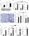
Loss of PLTP expression in the lung leads to a proinflammatory state. A) Lungs from A/J mice were examined for PLTP gene and protein expression and BALF PLTP activity following intranasal administration of negative control or PLTP siRNA. RQ, relative quantification vs. siRNA-treated mice. B) A significant increase in inflammatory cells was measured in the BALF of LPS-challenged mice that were administered PLTP siRNA. C) Comparative histological images of the 4 mouse groups. Scale bars = 100 μM. D) ERK and NF-κB activation were determined in whole lung tissue using multiplex antibodies for the phosphorylated and total forms of each protein. E) IFN-γ, TNF-α, and MMP-9 levels were determined in BALF by multiplex analysis and IL-1β levels by qPCR. RQ, quantification vs. vehicle- and control siRNA-treated mice. Graphs represent means ± sem.
Figure 7.
Figure 7.
Administration of PLTP protein prevents LPS-induced ERK and NF-κB activation and cytokine production in mouse lungs. A/J mice were intranasally administered LPS in combination with albumin or PLTP protein. A) A significant decrease in inflammatory cells was measured in the BALF of LPS-challenged mice that were administered PLTP protein. B) Comparative histological images of the 4 mouse groups. Scale bars = 100 μM. C) ERK and NF-κB activation was determined in whole lung tissue using multiplex antibodies for the phosphorylated and total forms of each protein. D) IFN-γ, TNF-α, and MMP-9 levels were determined in BALF by multiplex and IL-1β levels by qPCR. RQ, relative quantification vs. vehicle- and albumin-treated mice. Graphs represent means ± se.

References

    1. Day J. R., Albers J. J., Lofton-Day C. E., Gilbert T. L., Ching A. F., Grant F. J., O'Hara P. J., Marcovina S. M., Adolphson J. L. (1994) Complete cDNA encoding human phospholipid transfer protein from human endothelial cells. J. Biol. Chem. 269, 9388–9391 - PubMed
    1. Oka T., Kujiraoka T., Ito M., Egashira T., Takahashi S., Nanjee M. N., Miller N. E., Metso J., Olkkonen V. M., Ehnholm C., Jauhiainen M., Hattori H. (2000) Distribution of phospholipid transfer protein in human plasma: presence of two forms of phospholipid transfer protein, one catalytically active and the other inactive. J. Lipid. Res. 41, 1651–1657 - PubMed
    1. Vuletic S., Jin L. W., Marcovina S. M., Peskind E. R., Moller T., Albers J. J. (2003) Widespread distribution of PLTP in human CNS: evidence for PLTP synthesis by glia and neurons, and increased levels in Alzheimer's disease. J. Lipid Res. 44, 1113–1123 - PubMed
    1. Desrumaux C., Risold P. Y., Schroeder H., Deckert V., Masson D., Athias A., Laplanche H., Le Guern N., Blache D., Jiang X. C., Tall A. R., Desor D., Lagrost L. (2005) Phospholipid transfer protein (PLTP) deficiency reduces brain vitamin E content and increases anxiety in mice. FASEB J. 19, 296–297 - PubMed
    1. Albers J. J., Vuletic S., Cheung M. C. (2012) Role of plasma phospholipid transfer protein in lipid and lipoprotein metabolism. Biochim. Biophys. Acta 1821, 345–357 - PMC - PubMed

Publication types

MeSH terms