Review
. 2014 Feb 15;28(4):305-16.
doi: 10.1101/gad.235473.113.

The R-spondin/Lgr5/Rnf43 module: regulator of Wnt signal strength

Affiliations
Review

The R-spondin/Lgr5/Rnf43 module: regulator of Wnt signal strength

Wim de Lau et al. Genes Dev. .

Abstract

Lgr5 was originally discovered as a common Wnt target gene in adult intestinal crypts and colon cancer. It was subsequently identified as an exquisite marker of multiple Wnt-driven adult stem cell types. Lgr5 and its homologs, Lgr4 and Lgr6, constitute the receptors for R-spondins, potent Wnt signal enhancers and stem cell growth factors. The Lgr5/R-spondin complex acts by neutralizing Rnf43 and Znrf3, two transmembrane E3 ligases that remove Wnt receptors from the stem cell surface. Rnf43/Znrf3 are themselves encoded by Wnt target genes and constitute a negative Wnt feedback loop. Thus, adult stem cells are controlled by an intricate interplay of potent Wnt agonists, antagonists, and anti-antagonists.

Keywords: Lgr5; Rnf43/Znrf3; Rspondin; Wnt signal regulation.


Figures

Figure 1.
Figure 1.
Architecture of the intestinal epithelium. The small intestinal lumen is lined with a specialized simple epithelium consisting of crypts and villi. Four types of differentiated epithelial cells cover these villi: the absorptive enterocytes, mucous-secreting goblet cells, hormone-secreting enteroendocrine cells, and tuft cells. The crypts of Lieberkühn, epithelial invasions into the underlying connective tissue, harbor Lgr5 stem cells and their progeny, the TA cells. Cycling Lgr5 stem cells are interspersed with terminally differentiated Paneth cells at crypt bottoms. Small numbers of noncycling secretory progenitors located near the crypt bottom have recently been demonstrated to represent a noncycling “reserve” stem cell population (Buczacki et al 2013).
Figure 2.
Figure 2.
Canonical Wnt signaling (Nusse 2012). In cells devoid of a Wnt signal, free cytoplasmic β-catenin is actively targeted for degradation. This is accomplished by two scaffolding proteins (Apc and Axin) that bind β-catenin. They reside in the so-called destruction complex. Two kinases (CkI and Gsk3β) present in the same destruction complex sequentially phosphorylate a set of highly conserved Ser and Thr residues of β-catenin. Phospho-modified β-catenin becomes a substrate of the ubiquitin E3 ligase β-Trcp and is subsequently degraded in proteasomes. In the absence of Wnt signaling, Groucho proteins determine the nuclear DNA-binding proteins of the Tcf/Lef family to act as transcriptional repressors of Wnt target genes. Secreted Wnt proteins (19 family members) can induce signaling by interacting with Wnt receptor complexes consisting of a member of the Frizzled family (10 members) and the low-density lipid receptor family members Lrp5 or Lrp6. Wnt binding inactivates the destruction complex. As a direct consequence, β-catenin accumulates in the cytoplasm and nucleus and binds to members of the Tcf/Lef family, converting these into transcriptional activators. The Wnt signaling pathway is regulated extensively at the receptor–ligand level (Cruciat and Niehrs 2013). Secreted Frizzled-related proteins (Sfrp and Frzb) and Wnt inhibitory factor (WIF) can bind Wnt directly to prevent activation of receptors. Other Wnt antagonists, DKK1 and Wise, inhibit by binding to the LRP coreceptor. Recently discovered additional stem cell-specific regulators of canonical Wnt signaling (including Lgr4–6; the R-spondins; Rnf43; and its paralog, Znrf3) are the subject of this review.
Figure 3.
Figure 3.
Schematic representation of the domain composition of Lgr1–8, R-spondin1–4, and Rnf43/Znrf3. The eight members of the Lgr family of GPCRs are characterized by the presence of a large ECD composed of LRRs. This domain is flanked by N-terminal and C-terminal caps and a C-terminal Hinge region connecting the LRR domain with the 7TM. They can be further subdivided into classes A, B, and C. The class A receptor family comprises Lgr1, Lgr2, and Lgr3, which are receptors for the glycoprotein hormones follicle-stimulating hormone, luteinizing hormone, and thyroid-stimulating hormone, respectively. Individual human A-type receptors contain nine LRRs and a relatively long Hinge domain. The class B receptor family consists of Lgr4, Lgr5, and Lgr6. The ECD consists of 17 LRRs, and their Hinge domain is of intermediate length. Lgr7 and Lgr8 constitute the class C receptor family. Both bind insulin-like peptide hormones. They exhibit a very short Hinge domain. They are distinguished by the presence of an LDLa N-terminal domain that is essential for signaling. R-spondin proteins harbor two Furin repeats, both required for Wnt signal enhancement. The Furin-1 domain interacts with RNF43/ZNRF3, while the Furin-2 domain binds Lgr4–6. The TSR-1 domain binds Syndecan-type cell surface receptors. RNF43 and ZNRF3 are homologous RING-type E3 ligases and are structurally related to Grail. Typically, these single-pass transmembrane proteins have an extracellular PA domain and a cytoplasmic RING domain. The E3 ubiquitin ligase activity exerted by the RING domains of Rnf43 and Znrf3 targets lysine residues in the 7TM loops of Frizzled receptors. Substrate recognition may be dependent on an interaction of their associated PA domain with the Frizzled–LRP5/6 Wnt receptor complex.
Figure 4.
Figure 4.
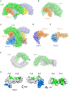
Crystal structures of the Lgr4/5, Rspo1/2, and Znrf3/Rnf43 complexes. (AF) Crystal structures of various complexes in cartoon and surface representation. (A) Lgr5–Rspo1 (Protein Data Bank [PDB] 4BSR) (Peng et al. 2013a). (B) Lgr4–Rspo1 (PDB 4KT1) (Wang et al. 2013a). (C) Lgr5–Rspo1–Rnf43 (PDB 4KNG) (Chen et al. 2013). (D) Znrf3–Rspo1 (PDB 4CDK) (Peng et al. 2013b). (E) Rnf43–Rspo2 (PDB 4C9V) (Zebisch et al. 2013). (F) Lgr4 (PDB 4LI1) (Xu et al. 2013). (G) Overview of the binding interfaces of Lgr5–Rspo1 and Rnf43–Rspo1, based on the crystal structure of Lgr5–Rspo1–Rnf43 (PDB 4KNG) (Chen et al. 2013). Shown are the contact footprints of Lgr5 on Rspo1 (in green) and Rnf43 on Rspo1 (in blue) in orthogonal views.
Figure 5.
Figure 5.
Regulation of Wnt receptor availability on stem cells. (Panels represent stills from animation that can be found at http://www.nymus3d.nl/video/rspondin.) (A) Upon binding of Wnt to the Fz/LRP receptor complex, some 100 Wnt target genes are activated, including Lgr5. Two other Wnt target genes, Rnf43 and Znrf3, encode transmembrane E3 ligases containing a cytoplasmic RING domain. These serve as components of a negative feedback loop. (B) Enzymatic interaction of the Rnf43/Znrf3-associated RING domain with the Fz/LRP complex leads to polyubiquitination of the intracellular loops of the 7TM domain of Fz. (C) The resulting endocytosis of Fz/Lrp abrogates Wnt signaling. The contribution of the PA domain in the specificity of this process is unclear. Whether Rnf43/Znrf3 are endocytosed with the Wnt receptor complex or segregate after their action is also unknown. When R-spondin is present (D), it is recruited to Lgr5 or its homolog, Lgr4 (E). This high-affinity interaction involves the Furin-2 repeat (Fu2) of R-spondin. This then enables the Furin-1 repeat (Fu1) of R-spondin to interact with Rnf43/Znrf3 (E), resulting in the membrane clearance of the latter (F). As a consequence, Wnt/Fz/Lrp receptor complexes persist on the plasma membrane, enhancing Wnt signal strength and duration.

Similar articles

Cited by

References

    1. Barker N, van Es JH, Kuipers J, Kujala P, van den Born M, Cozijnsen M, Haegebarth A, Korving J, Begthel H, Peters PJ, et al. 2007. Identification of stem cells in small intestine and colon by marker gene Lgr5. Nature 449: 1003–1007 - PubMed
    1. Barker N, Huch M, Kujala P, van de Wetering M, Snippert HJ, van Es JH, Sato T, Stange DE, Begthel H, van den Born M, et al. 2010. Lgr5(+ve) stem cells drive self-renewal in the stomach and build long-lived gastric units in vitro. Cell Stem Cell 6: 25–36 - PubMed
    1. Barker N, Rookmaaker MB, Kujala P, Ng A, Leushacke M, Snippert H, van de Wetering M, Tan S, van Es JH, Huch M et al. 2012. Lgr5(+ve) stem/progenitor cells contribute to nephron formation during kidney development. Cell Reports 2: 540–552 - PubMed
    1. Bell SM, Schreiner CM, Wert SE, Mucenski ML, Scott WJ, Whitsett JA 2008. R-spondin 2 is required for normal laryngeal-tracheal, lung and limb morphogenesis. Development 135: 1049–1058 - PubMed
    1. Binnerts ME, Kim KA, Bright JM, Patel SM, Tran K, Zhou M, Leung JM, Liu Y, Lomas WE 3rd, Dixon M, et al. 2007. R-Spondin1 regulates Wnt signaling by inhibiting internalization of LRP6. Proc Natl Acad Sci 104: 14700–14705 - PMC - PubMed

Publication types

MeSH terms

Substances