

Macular abnormalities in Italian patients with retinitis pigmentosa
- PMID: 24532797
- PMCID: PMC4078675
- DOI: 10.1136/bjophthalmol-2013-304082
Macular abnormalities in Italian patients with retinitis pigmentosa
Abstract
Aim: To investigate the prevalence of macular abnormalities in a large Caucasian cohort of patients affected by retinitis pigmentosa (RP).
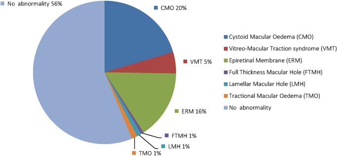
Methods: A retrospective study was performed by reviewing the medical records and optical coherence tomography (OCT) scans in a cohort of 581 RP patients in order to assess the presence of macular abnormalities -that is, cystoid macular oedema (CMO), epiretinal membrane (ERM), vitreo-macular traction syndrome, and macular hole.
Results: Macular abnormalities were observed in 524 (45.1%) out of the 1161 examined eyes. The most frequent abnormality was CMO, observed in 237 eyes (20.4%) from 133 patients (22.9%), followed by ERM, assessed in 181 eyes (15.6%) from 115 patients (19.8%). Moreover, vitreo-retinal abnormalities were significantly (p<0.05) associated with older age, cataract surgery, or cataract. CMO appeared to be significantly (p<0.05) associated with female gender, autosomic dominant inheritance pattern, and cataract.
Conclusions: Macular abnormalities are more frequent in RP compared to the general population. For that reason, screening RP patients with OCT is highly recommended to follow-up the patients, evaluate the natural history of disease, and identify those patients who could benefit from current or innovative therapeutic strategies.
Keywords: Diagnostic tests/Investigation; Dystrophy; Epidemiology; Macula; Retina.
Published by the BMJ Publishing Group Limited. For permission to use (where not already granted under a licence) please go to http://group.bmj.com/group/rights-licensing/permissions.
Figures

References
-
- Hagiwara A, Yamamoto S, Ogata K, et al. Macular abnormalities in patients with retinitis pigmentosa: prevalence on OCT examination and outcomes of vitreoretinal surgery. Acta Ophthalmol 2011;89:122–5 - PubMed
-
- Fishman GA, Fishman M, Maggiano J. Macular lesions associated with retinitis pigmentosa. Arch Ophthalmol 1977;95:798–803 - PubMed
-
- Giusti C, Forte R, Vingolo EM. Clinical pathogenesis of macular holes in patients affected by retinitis pigmentosa. Eur Rev Med Pharmacol Sci 2002;6:45–8 - PubMed
-
- Fishman GA. Retinitis pigmentosa. Genetic percentages. Arch Ophthalmol 1978;96:822–6 - PubMed
MeSH terms
LinkOut - more resources
Full Text Sources
Other Literature Sources