

The acute transcriptomic and proteomic response of HC-04 hepatoma cells to hepatocyte growth factor and its implications for Plasmodium falciparum sporozoite invasion
- PMID: 24532842
- PMCID: PMC4014276
- DOI: 10.1074/mcp.M113.035584
The acute transcriptomic and proteomic response of HC-04 hepatoma cells to hepatocyte growth factor and its implications for Plasmodium falciparum sporozoite invasion
Abstract
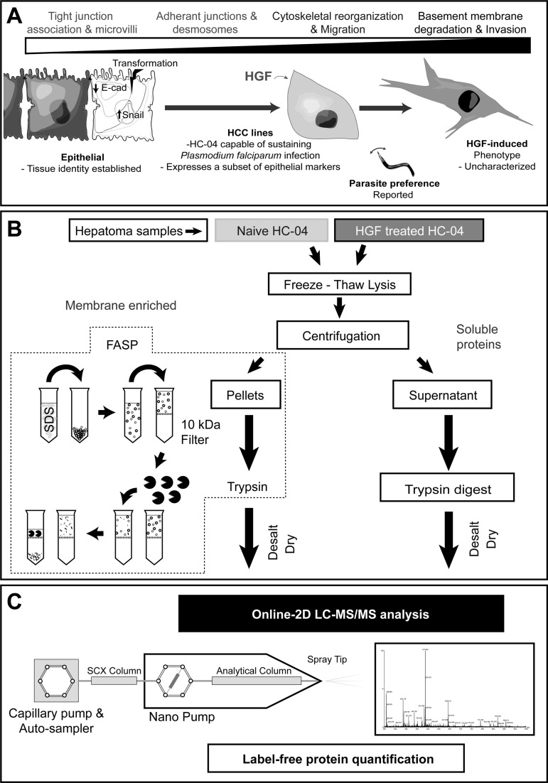
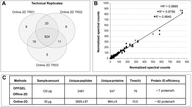
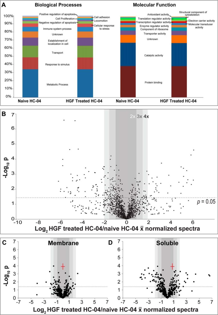
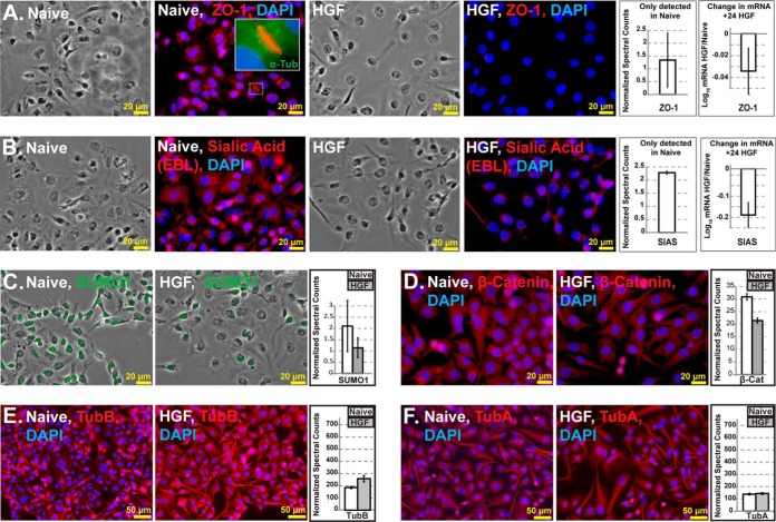
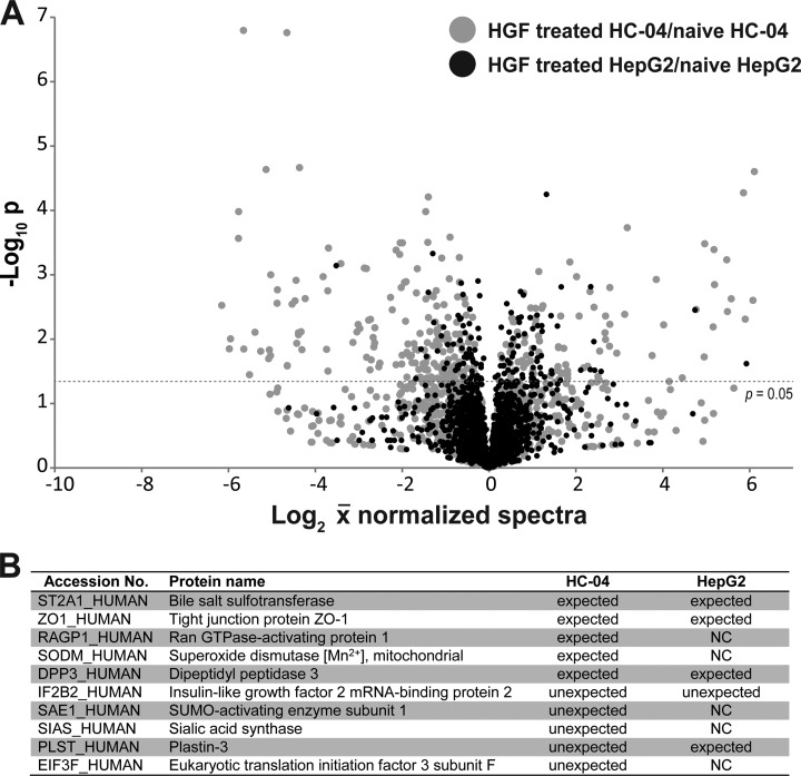
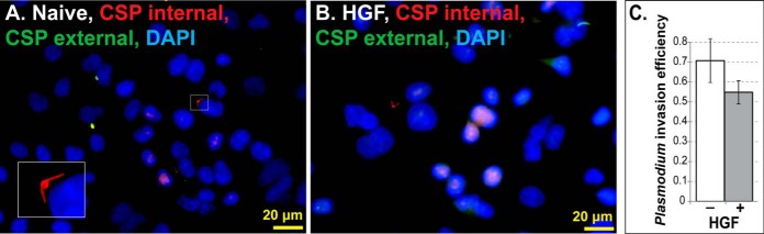
The routine study of human malaria liver-stage biology in vitro is hampered by low infection efficiency of human hepatocellular carcinoma (HCC) lines (<0.1%), poor understanding of steady-state HCC biology, and lack of appropriate tools for trace sample analysis. HC-04 is the only HCC that supports complete development of human malaria parasites. We hypothesized that HCCs are in various intermediate stages of the epithelial-mesenchymal transition (EMT) and HC-04s retain epithelial characteristics that permit infection. We developed a facile analytical approach to test this hypothesis viz. the HC-04 response to hepatocyte growth factor (HGF). We used online two-dimensional liquid chromatography tandem mass spectrometry (2D-LC-MS/MS) to quantify protein expression profiles in HC-04 pre-/post-HGF treatment and validated these results by RT-qPCR and microscopy. We successfully increased protein identification efficiency over offline-2D methods by 12-fold, using less sample material, allowing robust protein quantification. We observed expected up-regulation and down-regulation of EMT protein markers in response to HGF, but also unexpected cellular responses. We also observed that HC-04 is generally more susceptible to HGF-mediated signaling than what was observed for HepG2, a widely used, but poor malaria liver stage-HCC model. Our analytical approach to understanding the basic biology of HC-04 helps us understand the factors that may influence its utility as a model for malaria liver-stage development. We observed that HC-04 treatment with HGF prior to the addition of Plasmodium falciparum sporozoites did not facilitate cell invasion, which suggests unlinking the effect of HGF on malaria liver stage development from hepatocyte invasion. Finally, our 2D-LC-MS/MS approach and broadly applicable experimental strategy should prove useful in the analysis of various hepatocyte-pathogen interactions, tumor progression, and early disease events.
Conflict of interest statement
Figures
Similar articles
-
Temperature shift and host cell contact up-regulate sporozoite expression of Plasmodium falciparum genes involved in hepatocyte infection.PLoS Pathog. 2008 Aug 8;4(8):e1000121. doi: 10.1371/journal.ppat.1000121. PLoS Pathog. 2008. PMID: 18688281 Free PMC article.
-
Establishment of a human hepatocyte line that supports in vitro development of the exo-erythrocytic stages of the malaria parasites Plasmodium falciparum and P. vivax.Am J Trop Med Hyg. 2006 May;74(5):708-15. Am J Trop Med Hyg. 2006. PMID: 16687667
-
Flow Cytometry Based Detection and Isolation of Plasmodium falciparum Liver Stages In Vitro.PLoS One. 2015 Jun 12;10(6):e0129623. doi: 10.1371/journal.pone.0129623. eCollection 2015. PLoS One. 2015. PMID: 26070149 Free PMC article.
-
Current Challenges in the Identification of Pre-Erythrocytic Malaria Vaccine Candidate Antigens.Front Immunol. 2020 Feb 21;11:190. doi: 10.3389/fimmu.2020.00190. eCollection 2020. Front Immunol. 2020. PMID: 32153565 Free PMC article. Review.
-
Invasion of mammalian host cells by Plasmodium sporozoites.Bioessays. 2002 Feb;24(2):149-56. doi: 10.1002/bies.10050. Bioessays. 2002. PMID: 11835279 Review.
Cited by
-
A screen for Plasmodium falciparum sporozoite surface protein binding to human hepatocyte surface receptors identifies novel host-pathogen interactions.Malar J. 2024 May 16;23(1):151. doi: 10.1186/s12936-024-04913-2. Malar J. 2024. PMID: 38755636 Free PMC article.
-
HGF Secreted by Activated Kupffer Cells Induces Apoptosis of Plasmodium-Infected Hepatocytes.Front Immunol. 2017 Feb 6;8:90. doi: 10.3389/fimmu.2017.00090. eCollection 2017. Front Immunol. 2017. PMID: 28220125 Free PMC article.
-
Paraquat-Mediated Oxidative Stress in Anopheles gambiae Mosquitoes Is Regulated by An Endoplasmic Reticulum (ER) Stress Response.Proteomes. 2018 Nov 12;6(4):47. doi: 10.3390/proteomes6040047. Proteomes. 2018. PMID: 30424486 Free PMC article.
-
Evaluation of the HC-04 cell line as an in vitro model for mechanistic assessment of changes in hepatic cytochrome P450 3A during adenovirus infection.Drug Metab Dispos. 2014 Jul;42(7):1191-201. doi: 10.1124/dmd.113.056663. Epub 2014 Apr 24. Drug Metab Dispos. 2014. PMID: 24764148 Free PMC article.
-
The Selection of a Hepatocyte Cell Line Susceptible to Plasmodium falciparum Sporozoite Invasion That Is Associated With Expression of Glypican-3.Front Microbiol. 2019 Feb 28;10:127. doi: 10.3389/fmicb.2019.00127. eCollection 2019. Front Microbiol. 2019. PMID: 30891005 Free PMC article.
References
-
- Grisham J. W. (2009) Organizational Principles of the Liver, in The Liver: Biology and Pathobiology, Fifth Edition (ed Arias I. M.), John Wiley & Sons, Ltd, Chichester, UK: doi: 10.1002/9780470747919.ch1 - DOI
-
- Wilkening S., Stahl F., Bader A. (2003) Comparison of primary human hepatocytes and hepatoma cell line Hepg2 with regard to their biotransformation properties. Drug Metab. Dispos. 31, 1035–1042 - PubMed
-
- Liu L., Cao Y., Chen C., Zhang X., McNabola A., Wilkie D., Wilhelm S., Lynch M., Carter C. (2006) Sorafenib blocks the RAF/MEK/ERK pathway, inhibits tumor angiogenesis, and induces tumor cell apoptosis in hepatocellular carcinoma model PLC/PRF/5. Cancer Res. 66, 11851–11858 - PubMed
-
- Rosenberg R., Wirtz R. A., Schneider I., Burge R. (1990) An estimation of the number of malaria sporozoites ejected by a feeding mosquito. Trans. R. Soc. Trop. Med. Hyg. 84, 209–212 - PubMed
Publication types
MeSH terms
Substances
Grants and funding
LinkOut - more resources
Full Text Sources
Other Literature Sources
Research Materials
