. 2013 Dec;8(12):1183-1199.
doi: 10.2217/fvl.13.112.

Adeno-associated virus structural biology as a tool in vector development

Affiliations

Adeno-associated virus structural biology as a tool in vector development

Lauren M Drouin et al. Future Virol. 2013 Dec.

Abstract

Adeno-associated viruses (AAVs) have become important therapeutic gene delivery vectors in recent years. However, there are challenges, including intractable tissues/cell types and pre-existing immune responses, which need to be overcome for full realization of this system. This review addresses strategies aimed at improving AAV efficacy in the clinic through the creation of hybrid vectors that display altered or more targeted specific tissue tropisms, while also escaping recognition from host-derived neutralizing antibodies. Characterization of these viruses with respect to serotypes contributing to their capsid, using available 3D structures, enables the identification of regions critical for particular tropism and antigenic phenotypes. Structural information also allows for rational design of vectors with specific targeted tropisms for improved therapeutic efficacy.

Keywords: adeno-associated virus; capsid structure; chimera; directed evolution; gene therapy; rational design; tissue tropism.


Figures

Figure 1
Figure 1. Adeno-associated virus genome organization
Two inverted terminal repeats flank the adeno-associated ssDNA genome, which encodes the rep, cap and aap genes. The rep gene encodes four nonstructural proteins: Rep78, Rep68, Rep 52 and Rep40. Three overlapping VP proteins (VP1, VP2 and VP3) are expressed from the cap gene. AAP is expressed from a nonconventional start site nested in the cap gene.
Figure 2
Figure 2. A simplified adeno-associated virus phylogram
The AAV serotypes are separated into five clades (A–F) and two clonal isolates based on VP1 amino acid sequence. ClustalW2 Phylogeny [111] was used to generate the rooted phylogenetic tree. Distance values are shown for each serotype and indicate the number of substitutions as a proportion of the length of the alignment.
Figure 3
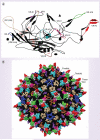
Figure 3. Adeno-associated virus capsid structure
(A) An adeno-associated virus (AAV) VP3 monomer is shown with a conserved core region consisting of eight antiparallel β-sheets (βB–βI) and an α-helix (αA). Loop insertions between the β-sheets vary among the AAV serotypes. Nine VRs (defined in [49]) are present on the capsid surface, and are color coded and denoted with roman numerals (I: purple; II: blue; III: yellow; IV: red; V: gray; VI: hot pink; VII: cyan; VIII: green; IX: brown; βHI loop: tan). (B) Locations of the VRs and viral asymmetric unit (shown in white) on the surface of the AAV capsid. The two-, three- and five-fold symmetry axes make up the border of the viral asymmetric unit, which is defined as the smallest repeating unit on the icosahedral capsid, and 60 of these compose the AAV capsid. It is indicated by a triangle on the surface of the AAV capsid, encompassing the area from the fivefold pore down to the twofold symmetry axis and outwards to the threefold protrusions. VR: Variable region.
Figure 4
Figure 4. Adeno-associated virus capsid interactions
(A) An adeno-associated virus capsid is composed of 60 VP monomers. A viral asymmetric unit is depicted (shown in white triangle), as demonstratd in Figure 3. (B) A space-filled surface representation of an adeno-associated virus monomer (reference monomer shown in green). (C) A dimer of monomers interacts to form a depression at the twofold axis of symmetry. (D) A trimer in which the interdigitation of three monomers forms the threefold spiky protrusions. (E) A pentamer with five interacting monomers, which has a characteristic central channel that spans the interior and exterior of the capsid.
Figure 5
Figure 5. Models of adeno-associated virus chimeras created by directed evolution
Exterior (left-hand side) and interior (right-hand side) capsid views of (A) adeno-associated virus (AAV)-DJ, (B) AAV-HAE1, (C) AAV-HAE2, (D) AAVM41 and (E) AAV-1829. Capsids are color coded by their contributing serotypes (and in lighter shades if there are multiple contributions from that serotype). AAV1: purple; AAV2: blue; AAV3: yellow; AAV4: red; AAV5: gray; AAV6: hot pink; AAV7: cyan; AAV8: green; AAV9: brown. The viral asymmetric unit is shown in white (as in Figure 3).
Figure 6
Figure 6. Models of adeno-associated virus chimeras created by rational capsid engineering
Exterior (left-hand side) and 2D surface roadmap views (right-hand side) of (A) adeno-associated virus (AAV2) Y/F mutants, (B) AAV2 Y/F and T/V quadruple mutant (Y444F + Y500F + Y730F + T491V), (C) AAV2 ↔ AAV8 loop variant (581–584 and 589–592), (D) AAV2i8, (E) AAV9 variant AAV9.45 and (F) AAV2.5. Capsids are colored gray and altered residues are color coded by their contributing serotypes (and in lighter shades if there are multiple contributions from that serotype). AAV1: purple; AAV2: blue; AAV3: yellow; AAV4: red; AAV5: gray; AAV6: hot pink; AAV7: cyan; AAV8: green; AAV9: brown. The viral asymmetric unit is shown in white on the left and black on the right (as in Figure 3).

References

    1. Mingozzi F, High KA. Therapeutic in vivo gene transfer for genetic disease using AAV: progress and challenges. Nat. Rev. Genet. 2011;12(5):341–355. - PubMed
    1. Yanez RJ, Porter AC. Therapeutic gene targeting. Gene Ther. 1998;5(2):149–159. - PubMed
    1. Peng KW. Strategies for targeting therapeutic gene delivery. Mol. Med. Today. 1999;5(10):448–453. - PubMed
    1. Raper SE, Chirmule N, Lee FS, et al. Fatal systemic inflammatory response syndrome in a ornithine transcarbamylase deficient patient following adenoviral gene transfer. 80. Mol. Genet. Metab. 2003;(1–2):148–158. - PubMed
    1. Hacein-Bey-Abina S, Garrigue A, Wang GP, et al. Insertional oncogenesis in 4 patients after retrovirus-mediated gene therapy of SCID-X1. J. Clin. Invest. 2008;118(9):3132–3142. - PMC - PubMed