

Sonic hedgehog improves redifferentiation of dedifferentiated chondrocytes for articular cartilage repair
- PMID: 24533105
- PMCID: PMC3922882
- DOI: 10.1371/journal.pone.0088550
Sonic hedgehog improves redifferentiation of dedifferentiated chondrocytes for articular cartilage repair
Abstract
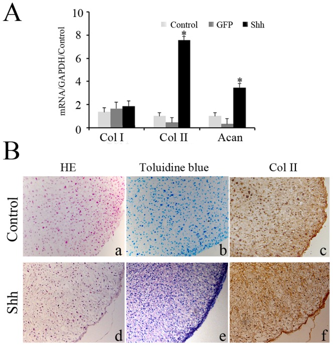
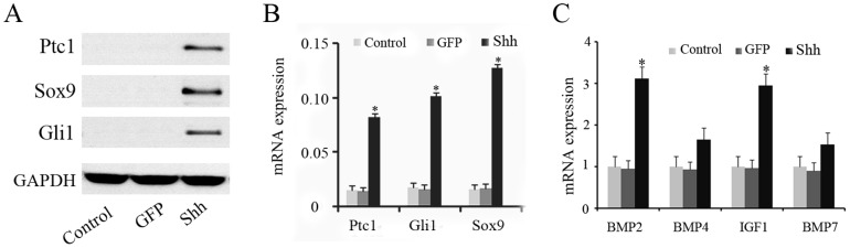
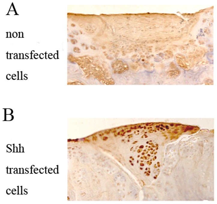
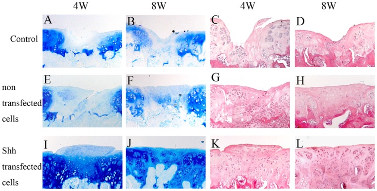
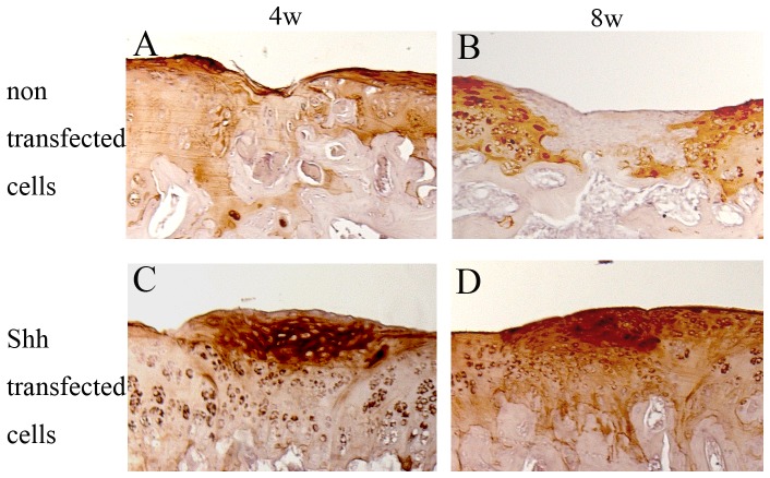
Sonic hedgehog (Shh) is involved in the induction of early cartilaginous differentiation of mesenchymal cells in the limb. We investigated whether Shh could promote redifferentiation of dedifferentiated chondrocytes and have a favorable effect on the regeneration of cartilage. Articular chondrocytes of rats were separated and cultured. The redifferentiation of dedifferentiated chondrocytes transfected with Shh was evaluated using monolayer and pellet culture system. The signaling molecules (Ptc 1, Gli 1 and Sox9) of the hedgehog pathway were investigated. A rat model of articular cartilage defect was used to evaluate cartilage repair after transplantation with dedifferentiated chondrocytes. After Shh gene transfer, the hedgehog pathway was upregulated in dedifferentiated chondrocytes. Real time-PCR and western blot analysis verified the stronger expression of Ptc1, Gli1 and Sox9 in Shh transfected cells. Shh upregulates the Shh signaling pathway and multiple cytokines (bone morphogenetic protein 2 and insulin-like growth factor 1) in dedifferentiated chondrocytes. After transplantation in the joint, histologic analysis of the regenerative tissues revealed that significantly better cartilage repair in rats transplanted with Shh transfected cells. These data suggest that Shh could induce redifferentiation of dedifferentiated chondrocytes through up-regulating Shh signaling pathway, and have considerable therapeutic potential in cartilage repair.
Conflict of interest statement
Figures
Similar articles
-
Targeted expression of SHH affects chondrocyte differentiation, growth plate organization, and Sox9 expression.J Bone Miner Res. 2004 Oct;19(10):1678-88. doi: 10.1359/JBMR.040706. Epub 2004 Jul 12. J Bone Miner Res. 2004. PMID: 15355563
-
Articular cartilage repair using dedifferentiated articular chondrocytes and bone morphogenetic protein 4 in a rabbit model of articular cartilage defects.Arthritis Rheum. 2008 Apr;58(4):1067-75. doi: 10.1002/art.23380. Arthritis Rheum. 2008. PMID: 18383381
-
Synergistic effects of Indian hedgehog and sonic hedgehog on chondrogenesis during cartilage repair.J Mol Histol. 2021 Apr;52(2):407-418. doi: 10.1007/s10735-021-09964-2. Epub 2021 Feb 17. J Mol Histol. 2021. PMID: 33598817
-
Regulation of senescence associated signaling mechanisms in chondrocytes for cartilage tissue regeneration.Osteoarthritis Cartilage. 2016 Feb;24(2):196-205. doi: 10.1016/j.joca.2015.07.008. Epub 2015 Jul 16. Osteoarthritis Cartilage. 2016. PMID: 26190795 Review.
-
[Research progress on signaling molecules involved in articular cartilage repair].Sheng Wu Yi Xue Gong Cheng Xue Za Zhi. 2019 Apr 25;36(2):343-348. doi: 10.7507/1001-5515.201806021. Sheng Wu Yi Xue Gong Cheng Xue Za Zhi. 2019. PMID: 31016955 Free PMC article. Review. Chinese.
Cited by
-
Hedgehog proteins and parathyroid hormone-related protein are involved in intervertebral disc maturation, degeneration, and calcification.JOR Spine. 2019 Nov 19;2(4):e1071. doi: 10.1002/jsp2.1071. eCollection 2019 Dec. JOR Spine. 2019. PMID: 31891120 Free PMC article.
-
MicroRNA-138 Inhibits Osteogenic Differentiation and Mineralization of Human Dedifferentiated Chondrocytes by Regulating RhoC and the Actin Cytoskeleton.JBMR Plus. 2018 Jul 18;3(2):e10071. doi: 10.1002/jbm4.10071. eCollection 2019 Feb. JBMR Plus. 2018. PMID: 30828688 Free PMC article.
-
Signaling pathways in cartilage repair.Int J Mol Sci. 2014 May 15;15(5):8667-98. doi: 10.3390/ijms15058667. Int J Mol Sci. 2014. PMID: 24837833 Free PMC article. Review.
-
Sonic hedgehog promotes chondrogenesis of rabbit bone marrow stem cells in a rotary cell culture system.BMC Dev Biol. 2019 Aug 12;19(1):18. doi: 10.1186/s12861-019-0198-4. BMC Dev Biol. 2019. PMID: 31401976 Free PMC article.
-
Strategies to Modulate the Redifferentiation of Chondrocytes.Front Bioeng Biotechnol. 2021 Nov 22;9:764193. doi: 10.3389/fbioe.2021.764193. eCollection 2021. Front Bioeng Biotechnol. 2021. PMID: 34881234 Free PMC article. Review.
References
-
- Benya PD, Shaffer JD (1982) Dedifferentiated chondrocytes reexpress the differentiated collagen phenotype when cultured in agarose gels. Cell 30: 215–224. - PubMed
-
- Hauselmann HJ, Fernandes RJ, Mok SS, Schmid TM, Block JA, et al. (1994) Phenotypic stability of bovine articular chondrocytes after long-term culture in alginate beads. J Cell Sci 107: 17–27. - PubMed
-
- Barbero A, Ploegert S, Heberer M, Martin I (2003) Plasticity of clonal populations of dedifferentiated adult human articular chondrocytes. Arthritis Rheum 48: 1315–1325. - PubMed
-
- Lin L, Zhou C, Wei X, Hou Y, Zhao L, et al. (2008) Articular cartilage repair using dedifferentiated articular chondrocytes and bone morphogenetic protein 4 in a rabbit model of articular cartilage defects. Arthritis Rheum 58: 1067–1078. - PubMed
Publication types
MeSH terms
Substances
LinkOut - more resources
Full Text Sources
Other Literature Sources
Research Materials
