

Vitreous incarceration in patients undergoing second 20-gauge pars plana vitrectomy for recurrent retinal detachment
- PMID: 24533182
- PMCID: PMC3912587
- DOI: 10.5402/2011/456191
Vitreous incarceration in patients undergoing second 20-gauge pars plana vitrectomy for recurrent retinal detachment
Abstract
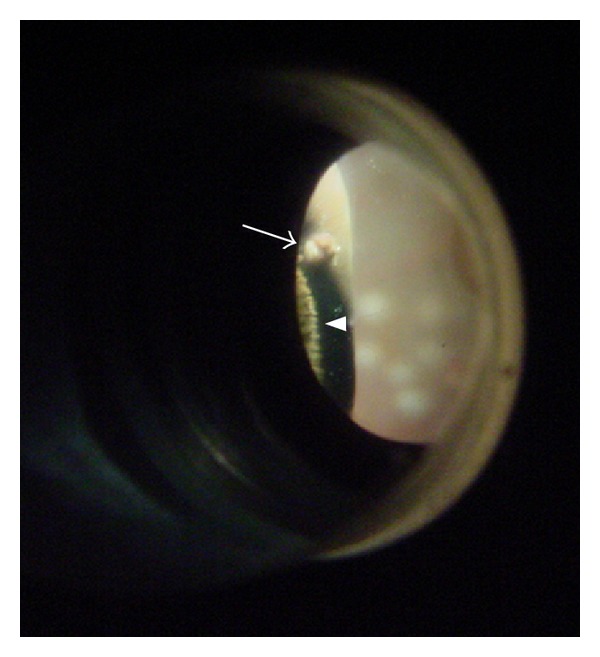
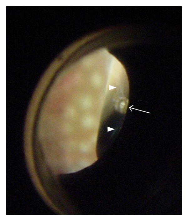
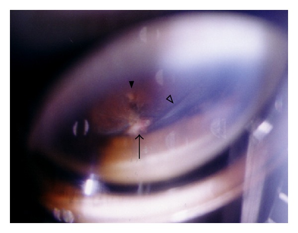
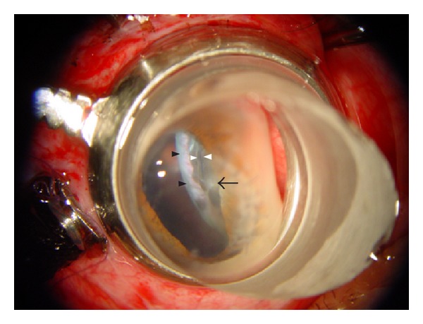
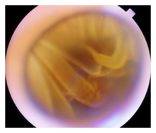
Purpose. To observe and classify vitreous incarcerations in patients undergoing second 20-gauge pars plana vitrectomy (PPV) for recurrent retinal detachment. Methods. Retrospective noncomparative consecutive case series. Eighty-two consecutive patients with recurrent retinal detachment were included. The previous sclerotomy sites were examined by our sclera depression method and the vitreous incarceration were classified into Grade 0-IV by their severity under surgical microscope before second surgery. The relationship of vitreous incarceration and different ports was statistically investigated in our included patients. Results. Vitreous incarceration in the previous sclerotomy sites were found frequently. Vitreous cutter sites were most involved, but the infusion pipe sites were the least. According to our classification and definition, Grade III and IV of vitreous incarceration in all the three different sclerotomy sites accounted for 32.5%. Grade II of vitreous incarceration consisted of 12.6%. Grade 0 and I in all the three different sclerotomy sites were 54.8%. The frequency of all grades of vitreous incarceration in light port or vitreous cutter port was significant higher than that in infusion port. Conclusions. Vitreous incarceration in light port and vitreous cutter port are found more common than in infusion port for 20-gauge PPV with our new method.
Figures
Similar articles
-
Ultrasound biomicroscopy of sclerotomy sites after pars plana vitrectomy for diabetic vitreous hemorrhage.Ophthalmology. 2000 Sep;107(9):1729-36. doi: 10.1016/s0161-6420(00)00213-x. Ophthalmology. 2000. PMID: 10964837
-
Fibrovascular ingrowth at sclerotomy sites in vitrectomized diabetic eyes with recurrent vitreous hemorrhage: ultrasound biomicroscopy findings.Ophthalmology. 2004 Jun;111(6):1215-21. doi: 10.1016/j.ophtha.2003.08.043. Ophthalmology. 2004. PMID: 15177974
-
Ultrasound biomicroscopy of sclerotomy sites: the effect of vitreous shaving around sclerotomy sites during pars plana vitrectomy.Retina. 2001;21(5):464-8. doi: 10.1097/00006982-200110000-00008. Retina. 2001. PMID: 11642375
-
Cryotherapy of the anterior retina and sclerotomy sites in diabetic vitrectomy to prevent recurrent vitreous hemorrhage: an ultrasound biomicroscopy study.Ophthalmology. 2005 Dec;112(12):2095-102. doi: 10.1016/j.ophtha.2005.07.010. Epub 2005 Oct 12. Ophthalmology. 2005. PMID: 16225926
-
[Dry transconjunctival sutureless 25-gauge vitrectomy in the treatment of pediatric cataract].Zhonghua Yan Ke Za Zhi. 2009 Aug;45(8):762-5. Zhonghua Yan Ke Za Zhi. 2009. PMID: 20021895 Review. Chinese.
References
-
- Lewis H, Aaberg TM, Abrams GW. Causes of failure after initial vitreoretinal surgery for severe proliferative vitreoretinopathy. American Journal of Ophthalmology. 1991;111(1):8–14. - PubMed
-
- Lewis H, Aaberg TM. Causes of failure after repeat vitreoretinal surgery for recurrent proliferative vitreoretinopathy. American Journal of Ophthalmology. 1991;111(1):15–19. - PubMed
-
- Lewis H, Aaberg TM. Anterior proliferative vitreoretinopathy. American Journal of Ophthalmology. 1988;105(3):277–284. - PubMed
-
- Elner SG, Elner VM, Diaz-Rohena R, Freeman HM, Tolentino FI, Albert DM. Anterior proliferative vitreoretinopathy: clinicopathologic, light microscopic and ultrastructural findings. Ophthalmology. 1988;95(10):1349–1357. - PubMed
LinkOut - more resources
Full Text Sources
Miscellaneous