

Influence of ulnar translation of the radial shaft in distal radius fracture on distal radioulnar joint instability
- PMID: 24533241
- PMCID: PMC3922857
- DOI: 10.1055/s-0033-1364093
Influence of ulnar translation of the radial shaft in distal radius fracture on distal radioulnar joint instability
Abstract
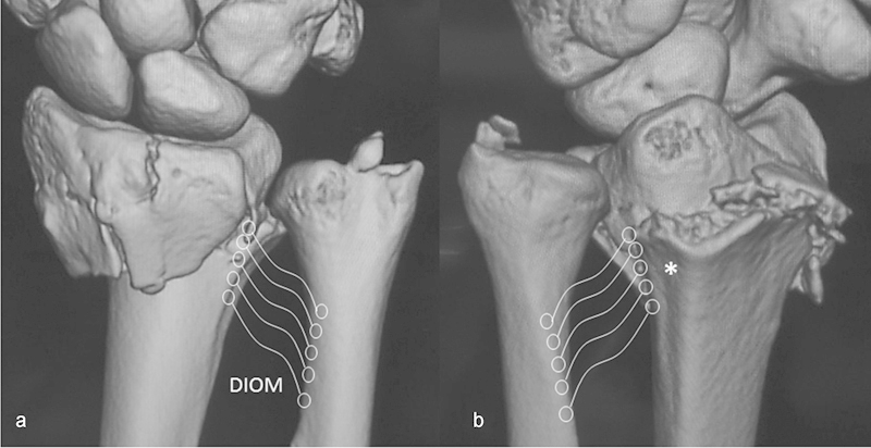
It has been reported that the distal interosseous membrane (DIOM) of the forearm constrains the dorsal dislocation of the distal radius. A residual ulnar translation deformity of the radial shaft in distal radius fractures has the potential to cause distal radioulnar joint (DRUJ) instability when triangular fibrocartilage complex (TFCC) injury is also present, because it may result in detensioning of the DIOM. Correction of ulnar translation of the radial shaft is critical because it restores DIOM tension, which then firmly holds the ulnar head in the concavity of the sigmoid notch.
Keywords: distal oblique bundle; distal radioulnar joint instability; distal radius fracture; interosseous membrane; translation.
Conflict of interest statement
Figures


References
-
- Kihara H, Short W H, Werner F W, Fortino M D, Palmer A K. The stabilizing mechanism of the distal radioulnar joint during pronation and supination. J Hand Surg Am. 1995;20(6):930–936. - PubMed
-
- Ward L D, Ambrose C G, Masson M V, Levaro F. The role of the distal radioulnar ligaments, interosseous membrane, and joint capsule in distal radioulnar joint stability. J Hand Surg Am. 2000;25(2):341–351. - PubMed
-
- Stuart P R, Berger R A, Linscheid R L, An K N. The dorsopalmar stability of the distal radioulnar joint. J Hand Surg Am. 2000;25(4):689–699. - PubMed
-
- Watanabe H, Berger R A, Berglund L J, Zobitz M E, An K N. Contribution of the interosseous membrane to distal radioulnar joint constraint. J Hand Surg Am. 2005;30(6):1164–1171. - PubMed
-
- Noda K, Goto A, Murase T, Sugamoto K, Yoshikawa H, Moritomo H. Interosseous membrane of the forearm: an anatomical study of ligament attachment locations. J Hand Surg Am. 2009;34(3):415–422. - PubMed
LinkOut - more resources
Full Text Sources
Other Literature Sources
