. 2014 Feb 18:10:42.
doi: 10.1186/1746-6148-10-42.

Development of a direct competitive ELISA for the detection of Mycoplasma bovis infection based on a monoclonal antibody of P48 protein

Affiliations

Development of a direct competitive ELISA for the detection of Mycoplasma bovis infection based on a monoclonal antibody of P48 protein

Ping Fu et al. BMC Vet Res. .

Abstract

Background: Mycoplasma bovis (M. bovis) is a major, but often overlooked, pathogen documented to cause respiratory disease, mastitis, and arthritis in cattle throughout China since 2008. Here, we report the development of a direct competitive enzyme-linked immunosorbent assay (Dc-ELISA) to detect M. bovis antibody.

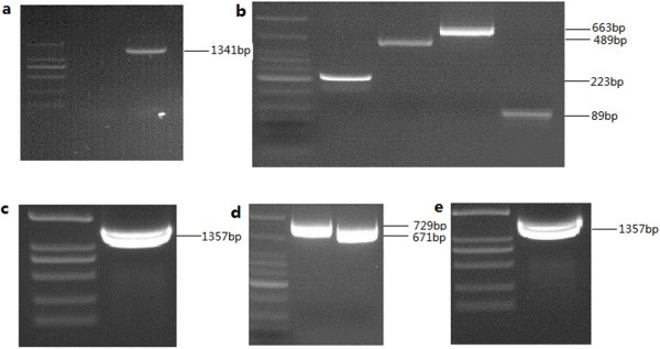
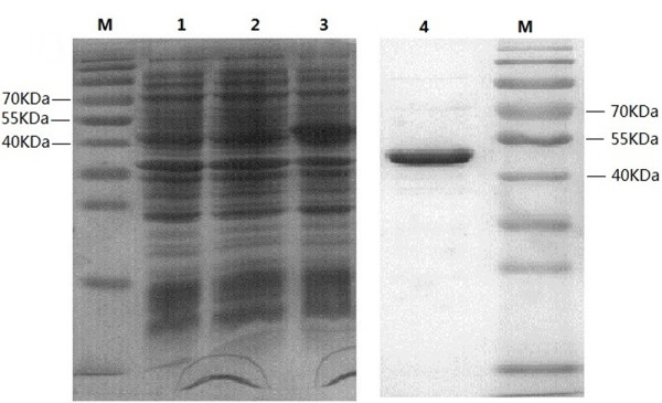
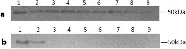
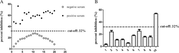
Results: We used a recombinant P48 protein and monoclonal antibody (mAb) 10E. MAb 10E, prepared against the recombinant P48 protein of M. bovis, identified all M. bovis strains with no cross-reactivity with other related pathogens. Coating micro plates with P48 protein instead of whole M. bovis cells as well as the use of mAb 10E produced a specific and sensitive Dc-ELISA for M. bovis antibody detection with a cut-off percent inhibition (PI) value of 32%. Compared with two commercial indirect ELISA (i-ELISA) kits, our Dc-ELISA offered a higher positive detection rate in 165 clinical bovine serum samples.

Conclusions: A rapid, sensitive, and reliable serological diagnosis method was developed for M. bovis, which can facilitate M. bovis surveillance, assisting researchers in understanding the ecology and epidemiology of M. bovis.


Figures

Figure 1
Figure 1
Identification and mutation of the P48 gene in M. bovis strain Hubei-1. a: Identification of P48 gene in M. bovis strain HB-1, using primer MB-F and MB-R primers. An amplified product of 1,341 bp as expected was obtained. b: Mutation of the bases on 192, 648, 1,275, 1,317 in the first PCR run. Four overlapping fragments, as expected, were 223 bp, 489 bp, 663 bp and 89 bp. d: Mutation of the base on 692 in the third PCR run, two overlapping fragments, 729 bp and 671 bp as expected were obtained. c and e: full length product obtained using purified fragments as template (MB-a and Mb-d’ primer) in the second and fourth PCR runs, respectively.
Figure 2
Figure 2
SDS-PAGE showing expression and purification of P48 in E. coli. All protein was subjected to 8-12% SDS-PAGE analysis and the gel was stained with Coomassie blue. M: prestained protein molecular weight marker (10-170 KDa), lane 1-3: total cell lysates of Transetta (DE3), Transetta (DE3) with PET28 (a+) plasmid, Transetta (DE3) with PET28 (a+)-P48 gene plasmid. Proteins were extracted after 1 mM IPTG induction for 6 h. Lane 4: purified P48 protein using His · Bind kits. Approximately 50 kDa protein band with a purity of 95% was observed.
Figure 3
Figure 3
Western blotting performed to investigate reactivity of mAb 10E with different pathogens. a: Lane 1-9 respectively was: purified P48 protein, M. bovis strain PG45, Hubei-1 SD-2, GY-2, PD-2, HRB-1, HY-1, HG-1. b: Lane 1-9 respectively was: purified P48 protein, M. bovis strain PG45, M. agalactiae, M. bovirhinis, M. arginie, IBRV, BADV3, BPIV3, BVDV.
Figure 4
Figure 4
Establishment of Dc-ELISA. (A) Distribution of inhibition values of 20 negative and 20 positive serum samples by Dc-ELISA. A cut-off value was set as 32% (mean + 2SD). Positive sera had PI values exceeding 32% and negative sera had PI values less than 32%. (B) Inhibition of binding of P48 protein-specific mAb 10E binding by antisera against various pathogenic species. Lanes 1-10 respectively are: BADV3, BVDV, BPIV3, BRSV positive control serum, Rabbit hyperimmune antisera against M. bovirhinis, M. arginine and M. agalactiae, rabbit negative serum, bovine negative M. bovis serum, bovine M. bovis positive serum. Only the M. bovis positive serum had clear inhibition of mAb binding. The Error bars indicate the standard deviations from three-well replications for each serum sample.

References

    1. Nicholas R, Ayling R. Mycoplasma bovis: disease, diagnosis, and control. Res Vet Sci. 2003;74(2):105. doi: 10.1016/S0034-5288(02)00155-8. - DOI - PubMed
    1. Xin J, Li Y, Guo D, Song N, Hu S, Chen C, Pei J, Cao P. First isolation of Mycoplasma bovis from calf lung with pneumoniae in China. Chin J Prev Vet Med. 2008;30(9):661–664.
    1. Qi J, Guo A, Cui P, Chen Y, Mustafa R, Ba X, Hu C, Bai Z, Chen X, Shi L. Comparative geno-plasticity analysis of Mycoplasma bovis HB0801 (Chinese isolate) PLoS ONE. 2012;7:8239. - PMC - PubMed
    1. Dénes B, Tenk M, Tekes L, Varga I, Ferenczné IP, Stipkovits L. Recognition of multiple Mycoplasma bovis antigens by monoclonal antibodies. Hybrid Hybridomics. 2003;22(1):11–16. doi: 10.1089/153685903321538035. - DOI - PubMed
    1. Ghadersohi A, Fayazi Z, Hirst R. Development of a monoclonal blocking ELISA for the detection of antibody to Mycoplasma bovis in dairy cattle and comparison to detection by PCR. Vet Immunol Immunopathol. 2005;104(3–4):183. - PubMed

Publication types

MeSH terms