Review
. 2014 Apr:27:136-43.
doi: 10.1016/j.ceb.2014.01.008. Epub 2014 Feb 17.

The role of protein dynamics in GPCR function: insights from the β2AR and rhodopsin

Affiliations
Review

The role of protein dynamics in GPCR function: insights from the β2AR and rhodopsin

Aashish Manglik et al. Curr Opin Cell Biol. 2014 Apr.

Abstract

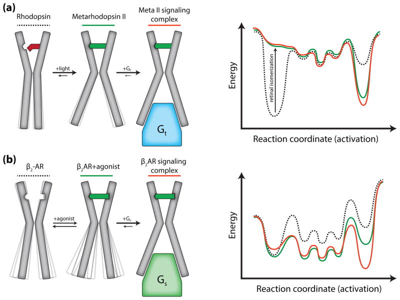
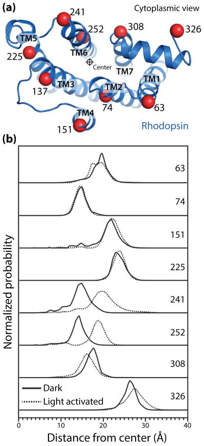
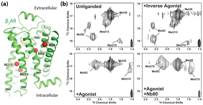
G protein-coupled receptors (GPCRs) are versatile signaling proteins that mediate complex cellular responses to hormones and neurotransmitters. Recent advances in GPCR crystallography have provided inactive and active state structures for rhodopsin and the β2 adrenergic receptor (β2AR). Although these structures suggest a two-state 'on-off' mechanism of receptor activation, other biophysical studies and observed signaling versatility suggest that GPCRs are highly dynamic and exist in a multitude of functionally distinct conformations. To fully understand how GPCRs work, we must characterize these conformations and determine how ligands affect their energetics and rates of interconversion. This brief review will compare and contrast the dynamic properties of rhodopsin and β2AR that shed light on the role of structural dynamics in their distinct signaling behaviors.


Figures

Figure 1
Figure 1
Differing models for activation of rhodopsin and the β2AR. (a) 11-cis-retinal bound rhodopsin is inactive with a cytoplasmic domain incapable of coupling to transducin (Gt). As the inactive conformation is the lowest energy state, dark rhodopsin displays minimal conformational heterogeneity. Light induced isomerization of the ligand to all-trans-retinal increases the energy of the inactive conformation resulting in a transition to the activated Meta II state and an opening of the cytoplasmic domain. The C-terminus of transducin interacts with the Meta II state to form the signaling complex. (b) Unliganded β2AR is conformationally dynamic as a result of smaller energetic differences between inactive, intermediate, and active states. Agonist binding increases β2AR dynamics by decreasing the energy of intermediate and active states. However, agonists do not fully stabilize the active state, and agonist bound β2AR primarily exists in inactive and intermediate conformations. Gs further stabilizes the active conformation and formation of the signaling complex is required for the receptor to completely transition to the active state.
Figure 2
Figure 2
Double electron-electron resonance (DEER) spectroscopy of rhodopsin activation (adapted from Altenbach et al. [42]). (a) View of the cytoplasmic surface of inactive rhodopsin. Nine sites, depicted with Cα carbons as red spheres, were site-specifically labeled pairwise with nitroxide spin probes. Distance distributions between these sites were determined for inactive and light-activated rhodopsin. (b) The displacement of each transmembrane helix from a central reference point is shown. Light activation of rhodopsin results in a 5 Å outward displacement of TM6, with smaller changes in TM1 and TM7. Notably, these data show that light activation results in a complete shift to the activated Meta II conformation in the absence of transducin, with almost no receptor in inactive or intermediate conformations.
Figure 3
Figure 3
13CH3ε-methionine NMR spectroscopy of β2AR (adapted from Nygaard et al. [44]). (a) View of the β2AR transmembrane helices showing 13CH3ε-methionine labeled carbons as red spheres. NMR peaks in HSQC spectra originate from four distinct sites. (b) HSQC spectra of β2AR in four states: unliganded, bound to inverse agonist carazolol, to agonist BI-167107, and to BI-167107 with the G protein mimetic nanobody Nb80.

Similar articles

Cited by

References

    1. Insel PA, Head BP, Ostrom RS, Patel HH, Swaney JS, Tang CM, Roth DM. Caveolae and lipid rafts: G protein-coupled receptor signaling microdomains in cardiac myocytes. Ann N Y Acad Sci. 2005;1047:166–172. - PubMed
    1. Shcherbakova OG, Hurt CM, Xiang Y, Dell’acqua ML, Zhang Q, Tsien RW, Kobilka BK. Organization of {beta}-adrenoceptor signaling compartments by sympathetic innervation of cardiac myocytes. J Cell Biol. 2007;176(4):521–533. - PMC - PubMed
    1. Kovacs JJ, Hara MR, Davenport CL, Kim J, Lefkowitz RJ. Arrestin development: Emerging roles for beta-arrestins in developmental signaling pathways. Dev Cell. 2009;17(4):443–458. - PMC - PubMed
    1. Xiang Y, Kobilka B. The pdz-binding motif of the beta2-adrenoceptor is essential for physiologic signaling and trafficking in cardiac myocytes. Proceedings of the National Academy of Sciences of the United States of America. 2003;100(19):10776–10781. - PMC - PubMed
    1. Irannejad R, Tomshine JC, Tomshine JR, Chevalier M, Mahoney JP, Steyaert J, Rasmussen SG, Sunahara RK, El-Samad H, Huang B, von Zastrow M. Conformational biosensors reveal gpcr signalling from endosomes. Nature. 2013;495(7442):534–538. - PMC - PubMed

Publication types