. 2014 May;4(5):564-77.
doi: 10.1158/2159-8290.CD-13-0419. Epub 2014 Feb 17.

NUP98-PHF23 is a chromatin-modifying oncoprotein that causes a wide array of leukemias sensitive to inhibition of PHD histone reader function

Affiliations

NUP98-PHF23 is a chromatin-modifying oncoprotein that causes a wide array of leukemias sensitive to inhibition of PHD histone reader function

Sheryl M Gough et al. Cancer Discov. 2014 May.

Abstract

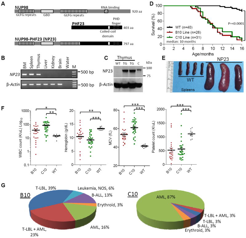
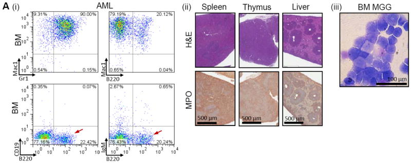
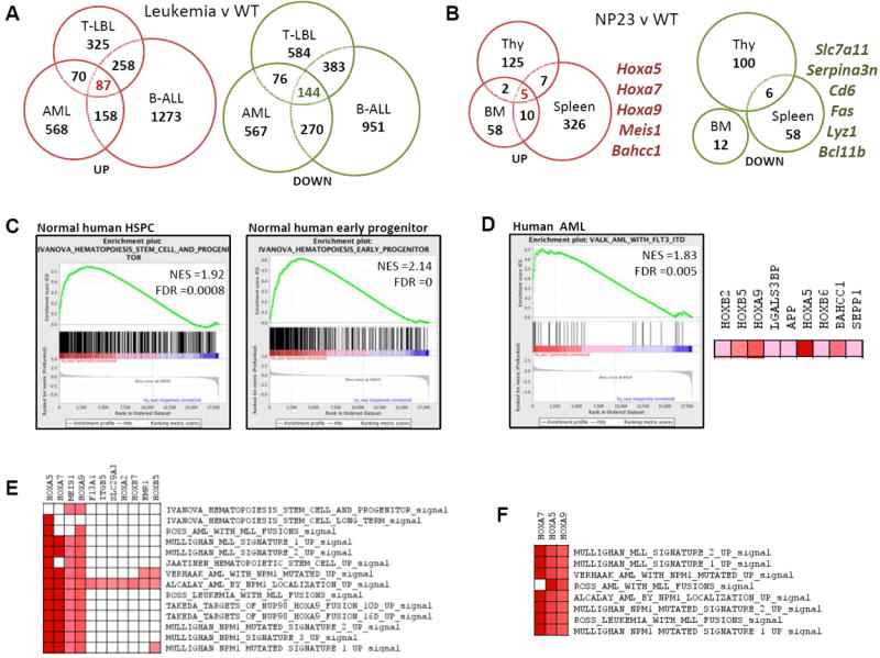
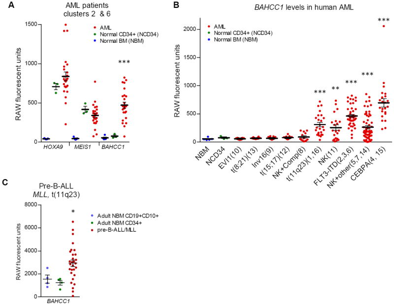
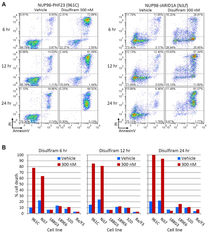
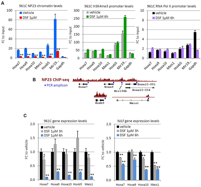
In this report, we show that expression of a NUP98-PHF23 (NP23) fusion, associated with acute myeloid leukemia (AML) in humans, leads to myeloid, erythroid, T-cell, and B-cell leukemia in mice. The leukemic and preleukemic tissues display a stem cell-like expression signature, including Hoxa, Hoxb, and Meis1 genes. The PHF23 plant homeodomain (PHD) motif is known to bind to H3K4me3 residues, and chromatin immunoprecipitation experiments demonstrated that the NP23 protein binds to chromatin at a specific subset of H3K4me3 sites, including at Hoxa, Hoxb, and Meis1. Treatment of NP23 cells with disulfiram, which inhibits the binding of PHD motifs to H3K4me3, rapidly and selectively killed NP23-expressing myeloblasts; cell death was preceded by decreased expression of Hoxa, Hoxb, and Meis1. Furthermore, AML driven by a related fusion gene, NUP98-JARID1A (NJL), was also sensitive to disulfiram. Thus, the NP23 mouse provides a platform to evaluate compounds that disrupt binding of oncogenic PHD proteins to H3K4me3.


Conflict of interest statement

CONFLICTS OF INTEREST STATEMENT: J.M.D. is a consultant to Sirtris-GlaxoSmithKline.

Figures

Figure 1
Figure 1. The NUP98-PHF23 (NP23) fusion protein is a multi-lineage oncoprotein
(A) Germline NUP98 (upper) and PHF23 (middle), and the aberrant NUP98-PHF23 fusion protein (lower). (B) Expression of the NP23 transgene in bone marrow (BM), spleen and thymus. (C) Immunoblot of NP23 protein in NP23 transgenic (TG) thymus. WT, wild type. C, positive control. (D) Survival curves of B10, C10 and WT littermate controls. (E) Representative necropsy images of splenomegaly in NP23 leukemia. (F) WBC, hemoglobin, MCV and platelet counts at disease presentation. Note the log10 scale on WBC graph. (G) Frequency of leukemia subtypes by founder line. Some mice found dead with evidence of leukemia could not be further evaluated and are designated “leukemia, not otherwise specified (NOS)”. T-test p values; *<0.05, **<0.01, ***<0.001.
Figure 2
Figure 2. NP23 AML
(A) AML from mouse 820; (i) FACS profiles show Mac1+/Gr1+ and Mac1+/B220dim cells in the bone marrow. The B220+ cells are CD19 and sIgM negative (red arrows) and are therefore not typical B-cells. (ii) H&E and MPO IHC of spleen, thymus and liver showing loss of normal organ architecture and infiltration of MPO+ malignant cells. (iii) May-Grunwald Giemsa (MGG) stained BM myeloblasts.
Figure 3
Figure 3. NP23 leukemias exhibit Hox/Meis1 stem cell-like gene expression signatures that are enriched in human HSPC and AML genesets
(A) Genes >1.5-fold overexpressed (red) or underexpressed (green) in AML, pre-T LBL and pre-B1 ALL compared to WT BM, thymus or spleen (Table S3). Eighty seven genes were overexpressed and 144 decreased independent of tissue type (Tables S4-S5). (B) Genes >1.5-fold overexpressed (red) or underexpressed (green) in NP23 BM, thymus or spleen compared to WT controls. Note a core set of only five genes are overexpressed irrespective of tissue type (Table S6). (C) Enrichment profiles of NP23 AML overexpressed genes in (A) human HSPC and early progenitor genesets, and (D) human AML with FLT3-ITD. Right, a subset of leading edge genes from the FLT3-ITD geneset. A subset of GSEA leading edge genes overexpressed in (E) NP23 AML and enriched in human HSPC, early progenitor and AML genesets; and (F) NP23 pre-T LBL and enriched in human leukemic genesets.
Figure 4
Figure 4. BAHCC1 expression in human AML and pre-B-ALL genesets
(A) Gene expression levels of HOXA9 (209905_at), MEIS1 (204069_at) and BAHCC1 (219218_at) in human AML, normal CD34+HSCs and normal bone marrow (GSE1159) (22). (B) BAHCC1 (219218_at) expression levels in human AMLs grouped by cytogenetic lesion (patient cluster#) (GSE1159) (22). NK, normal karyotype. Comp, complex karyotype. Other, misc chromosomal aberrations. (C) BAHCC1 expression in human pre-B-ALL with MLL gene rearrangement (GSE33315) (23). T-test p values are relative to normal CD34+ (HSC) samples in (A) and (B), and relative to adult NBM CD19+CD10+ precursor B-cells in (C); *<0.05, **<0.01, ***<0.001.
Figure 5
Figure 5. ChIP-seq identifies NP23-specific H3K4me3 enrichment and direct NP23 targets genome-wide
(A) H3K4me3 enriched genes in two NP23 pre-T LBL derived cell lines 106A and 748T, compared to H3K4me3 enriched genes in a non-NP23 control pre-T LBL cell line (murine SCL-LMO1, 7298/2). 429 sites were specific to NP23 106A and 748T (Table S7). (B) Overlap of NP23 T-cell H3K4me3 enriched genes and genes overexpressed in pre-T LBL. (C) H3K4me3 and H3K27me3 ChIP-seq at the 5’ Hoxa locus in 106A and 748T, compared to control cell line 7298/2. (D) H3K4me3, H3K27me3 and NP23 ChIP-seq at the 5’ Hoxa locus of NP23 AML-derived myeloblastic cell line 961C. (E) NP23 targets only 160 (1.6%) of the 9863 H3K4me3 enriched sites at annotated genes genome-wide (Table S8). (F) Comparison of genes with dual NP23 and H3K4me3 enrichment, to genes overexpressed in AML and healthy NP23 BM (Table S9). Hoxa7, Hoxa9, Meis1 and Hoxb5 are overexpressed in both healthy NP23 BM and NP23 AML, and are direct targets of NP23. H3K4me3, H3K27me3 and NP23 ChIP-seq at Meis1 (G), the Hoxb locus (H), and at Bahcc1. Note, Bahcc1 is void of NP23.
Figure 6
Figure 6. Disulfiram selectively kills leukemic cells driven by H3K4me3 binding NUP98-PHD fusions
(A) Apoptotic (PI+/AnnexinV+) analysis of NUP98-PHF23 (961C) and NUP98-JARID1A (NJLf) AML cell lines at 6, 12 and 24 hours following treatment with 300 nM disulfiram or vehicle (DMSO). (B) Graphed proportions of apoptotic/dead cells shown in (A). Early apoptotic, late apoptotic and necrotic cell populations are combined to represent “% cell death” in NUP98-PHD cell lines 961C and NJLf, compared to AML control (non-NP23) cell lines 188G3 (NUP98-HOXD13), 189E6 (NUP98-HOXD13), 32D and Ba/F3. Representative of three independent experiments.
Figure 7
Figure 7. Disulfiram treatment leads to reduced NP23 chromatin binding and target gene expression
(A) ChIP of promoter-associated NP23 fusion protein at target loci and Gapdh in 961C cells after 6 hours 1μM disulfiram (DSF) treatment. NP23 (left) H3K4me3 (middle) and RNA polymerase II (right) levels. (B) ChIP Q-PCR amplicon locations (blue square) are indicated relative to target gene and NP23 ChIP-seq tracks (as shown in Fig. 5). (C) RQ-PCR quantification of mRNA transcripts of target genes Hoxa7, Hoxa9, Hoxa10, Hoxb5 (not expressed in NJLf) and Meis1, in 961C (left) and NJLf (right) cells treated with disulfiram compared to vehicle (DMSO) treated cells. Ct values were normalized to internal ribosomal controls. P values are relative to vehicle control. Error bars indicate SEM; T-test P values are indicated; p *<0.05, **<0.01.

References

    1. Gough SM, Slape CI, Aplan PD. NUP98 gene fusions and hematopoietic malignancies: common themes and new biological insights. Blood. 2011;118:6247–57. - PMC - PubMed
    1. Lisboa S, Cerveira N, Bizarro S, Correia C, Vieira J, Torres L, et al. POU1F1 is a novel fusion partner of NUP98 in acute myeloid leukemia with t(3;11)(p11;p15) Mol Cancer. 2013;12:5. - PMC - PubMed
    1. Hollink IH, van den Heuvel-Eibrink MM, Arentsen-Peters ST, Pratcorona M, Abbas S, Kuipers JE, et al. NUP98/NSD1 characterizes a novel poor prognostic group in acute myeloid leukemia with a distinct HOX gene expression pattern. Blood. 2011;118:3645–56. - PubMed
    1. Pineault N, Buske C, Feuring-Buske M, Abramovich C, Rosten P, Hogge DE, et al. Induction of acute myeloid leukemia in mice by the human leukemia-specific fusion gene NUP98-HOXD13 in concert with Meis1. Blood. 2003;101:4529–38. - PubMed
    1. Wang GG, Cai L, Pasillas MP, Kamps MP. NUP98-NSD1 links H3K36 methylation to Hox-A gene activation and leukaemogenesis. Nat Cell Biol. 2007;9:804–12. - PubMed

Publication types

MeSH terms

Associated data