. 2014 Feb 17;204(4):507-22.
doi: 10.1083/jcb.201310083.

CDK phosphorylation of SLD-2 is required for replication initiation and germline development in C. elegans

Affiliations

CDK phosphorylation of SLD-2 is required for replication initiation and germline development in C. elegans

Vincent Gaggioli et al. J Cell Biol. .

Erratum in

  • J Cell Biol. 2014 Mar 17;204(6):1075

Abstract

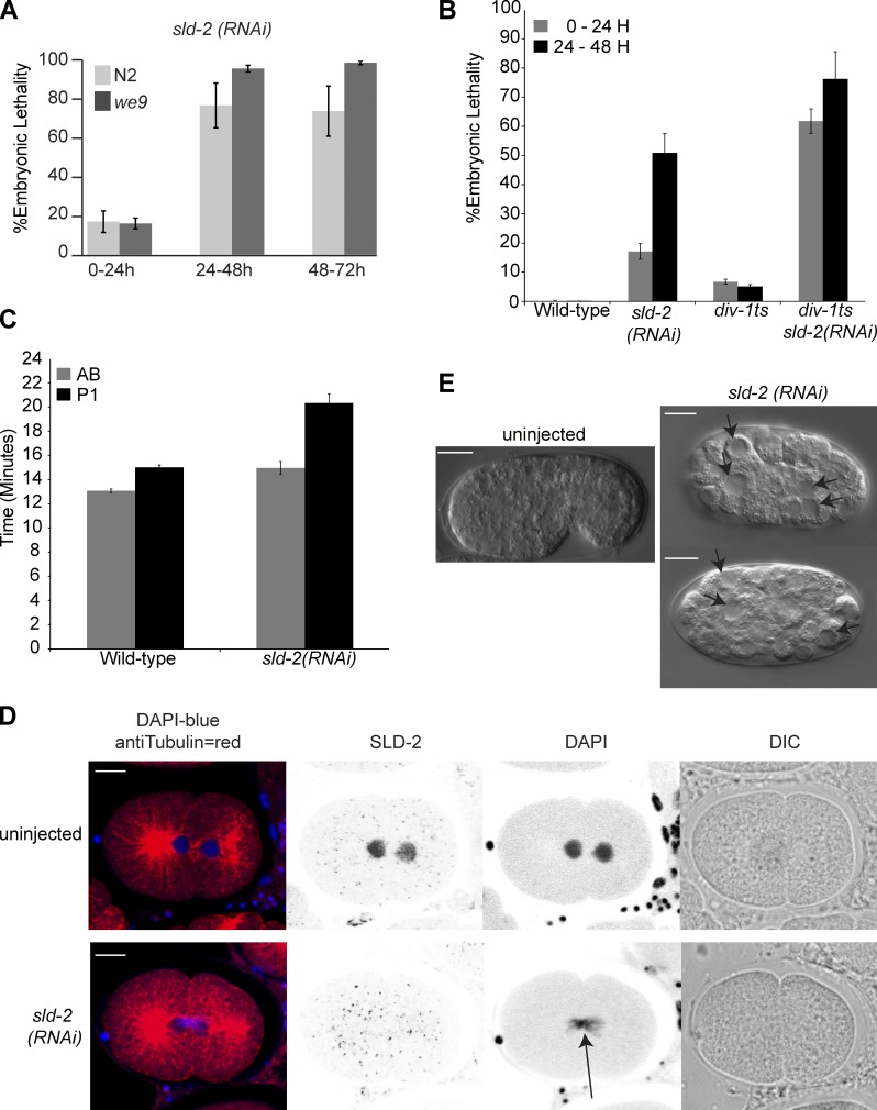
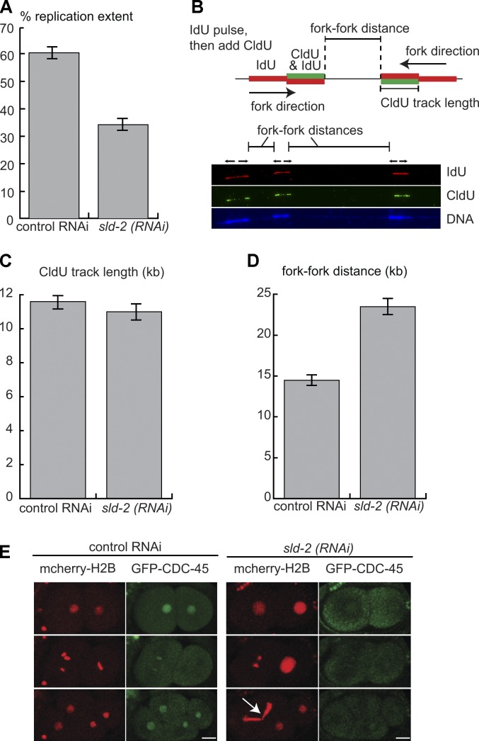
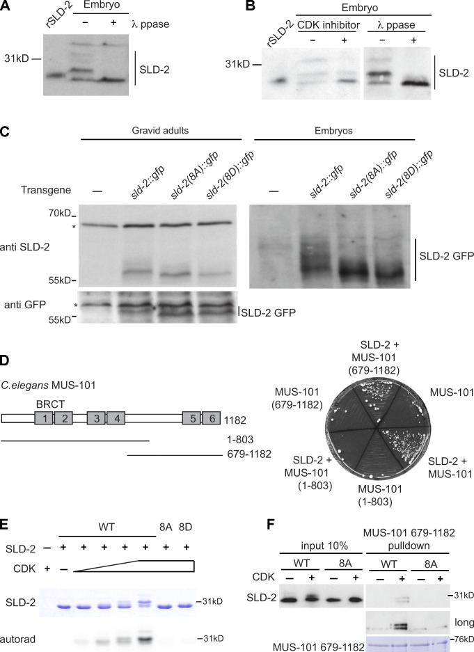
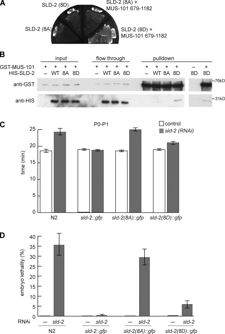
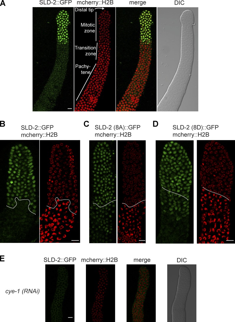
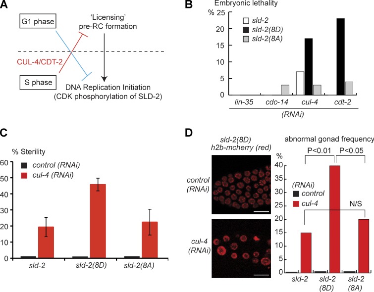
Cyclin-dependent kinase (CDK) plays a vital role in proliferation control across eukaryotes. Despite this, how CDK mediates cell cycle and developmental transitions in metazoa is poorly understood. In this paper, we identify orthologues of Sld2, a CDK target that is important for DNA replication in yeast, and characterize SLD-2 in the nematode worm Caenorhabditis elegans. We demonstrate that SLD-2 is required for replication initiation and the nuclear retention of a critical component of the replicative helicase CDC-45 in embryos. SLD-2 is a CDK target in vivo, and phosphorylation regulates the interaction with another replication factor, MUS-101. By mutation of the CDK sites in sld-2, we show that CDK phosphorylation of SLD-2 is essential in C. elegans. Finally, using a phosphomimicking sld-2 mutant, we demonstrate that timely CDK phosphorylation of SLD-2 is an important control mechanism to allow normal proliferation in the germline. These results determine an essential function of CDK in metazoa and identify a developmental role for regulated SLD-2 phosphorylation.


Figures

Figure 1.
Figure 1.
Bioinformatic screen for novel Sld2 orthologues. (A) Scale diagram of the two regions of homology (I [blue] and II [green]) between Sld2 from budding yeast and human RecQL4. (B) Alignment of homology region I between two yeast Sld2 proteins and three Sld2-RecQ4 proteins (see Table S1 for accession numbers). The first residue is the start methionine of these proteins. Residues with the greatest identity are highlighted in yellow, and regions of similarity are highlighted in pink. The position of the sld2-5 mutation and residues important (*) or unimportant (#) for Sld2 function in budding yeast are indicated above. (C) Alignment of Sld2 homology region I of some of the newly identified Sld2/RecQ4 orthologues as in B. The first residue is the start methionine, or the number of amino acids away from the first methionine is given in brackets. (D) Representation of Sld2/RecQ4 protein family as a phylogenetic tree. This tree is unrooted and not to scale. Multiple orthologues were found for each phylum (Table S1), but for each branch, only a single species is represented. H. sapiens, Homo sapiens; O. tauri, Ostreococcus tauri; M. brevicollis, Monosiga brevicollis; T. adhaerens, Trichoplax adhaerens; C. intestinalis, Ciona intestinalis; A. queenslandica, Amphimedon queenslandica; R. oryzae, Rhizopus oryzae; E. siliculosus, Ectocarpus siliculosus.
Figure 2.
Figure 2.
C. elegans sld-2 is essential. (A) Mean embryonic lethality after sld-2(RNAi) by injection into young adult N2 or RNAi-sensitive we9 worms at 15°C (n = 11). Embryo lethality is scored as the number of unhatched eggs relative to the total number of F1 eggs. (B) Mean embryonic lethality for wild-type N2 and div-1(or148ts) worms with and without sld-2(RNAi) injection at 21.5°C, 0–24 and 24–48 h after egg laying. n = 20 except for sld-2(RNAi) in which n = 51. (C) The timing of cell division in early embryos, extruded from N2 worms with or without injection of sld-2 RNAi, was analyzed by time-lapse differential interference contrast (DIC) imaging. The lengths of time after P0 cell nuclear envelope breakdown until the onset of nuclear envelope breakdown in the AB blastomere or the P1 blastomere are shown. n = 9. (D) Images of the early C. elegans embryo from we9 worms with or without sld-2 (RNAi) by injection. SLD-2 was visualized by anti–SLD-2 immunofluorescence using affinity-purified Ab 5058. An arrow indicates DNA stuck in the plane of cytokinesis—the cut phenotype. (E) Differential interference contrast images showing C. elegans embryos of the same age, from mothers of the we9 strain either uninjected or injected with sld-2 (RNAi). Arrows indicate large undifferentiated cells. Error bars are SEMs. Bars, 10 µM.
Figure 3.
Figure 3.
C. elegans sld-2 is required for replication initiation. (A) Replication extent of DNA fibers from control or sld-2 RNAi embryos from the we9 worm strain. Chitinase-treated embryos from synchronized adults were pulse labeled by 20-min incubation with IdU followed by DNA isolation and stretching onto silanized slides. Replicating DNA was visualized by anti-IdU immunofluorescence, and replication extent was calculated by dividing the summed lengths of IdU tracks on a single fiber by the total length of the fiber. n = 53. (B) Double pulse labeling of replicating embryonic cells allows the discrimination of replication initiation and elongation events. Chitinase-treated embryos are first pulse labeled with IdU (red) and then CldU (green). Tracks that are red followed by red/green are elongating forks, and the green track length provides a measure of fork rate. Gaps between two red/green tracks indicate interfork distances. (bottom) Example of a labeled C. elegans embryo DNA fiber. The DNA label cross reacts with the IdU staining, which makes the DNA labeling more intense over IdU tracks. (C) CldU track length in double pulse-labeled embryos isolated from control (n = 89) or sld-2 RNAi (n = 55) we9 worms after feeding. (D) Mean fork–fork distances from the experiment in C. For control RNAi, n = 116; for sld-2 RNAi by feeding, n = 98. (E) Visualization of the replicative helicase component CDC-45 in worm embryos extruded from mothers either injected with buffer (control) or with sld-2 (RNAi) 36 h after injection. The arrow indicates DNA stuck in the plane of cytokinesis—the cut phenotype. Error bars are SEMs. Bars, 10 µM.
Figure 4.
Figure 4.
C. elegans SLD-2 is CDK phosphorylated in vivo and binds to MUS-101 in a phosphodependent manner. (A) Chitinase-treated embryos were treated with or without λ phosphatase (ppase) for 30 min at 25°C and analyzed by anti–SLD-2 (Ab 5058) Western blotting. Recombinant, E. coli–expressed SLD-2 (rSLD-2) has slightly lower mobility than endogenous, dephosphorylated SLD-2 because it is 6HIS tagged. (B) As in A, Chitinase-treated embryos were treated with or without CDK inhibitor III for 30 min at 25°C. A different exposure of the right-hand blot is also shown in Fig. S2 A. (C) Anti–SLD-2 (top) or anti-GFP Western (bottom) of SLD-2::GFP expressed from the indicated transgenes in gravid adult extracts (left) and embryo extracts (right). Asterisks mark nonspecific bands. The 8A or 8D sld-2 alleles correspond to mutants with all eight CDK consensus sites mutated to alanine or aspartic acid, respectively. (D, left) Diagram of C. elegans MUS-101 showing the position of the six BRCT repeats. (right) Selective growth medium after yeast two-hybrid analysis between MUS-101 (bait) and SLD-2 (prey). (E) Coomassie stain (top) and autoradiogram (bottom) of in vitro CDK phosphorylation of recombinant T7/6HIS-tagged SLD-2. (F) GST pull-down with GST–MUS-101 (679–1,182) and T7/6HIS SLD-2. (top) An anti-T7 Western of recombinant SLD-2. The 10% input and the short exposure are equal exposure times. (bottom) Coomassie stain of GST MUS-101 after the pull-down. WT, wild type.
Figure 5.
Figure 5.
C. elegans SLD-2 is an essential CDK target. (A) Selective growth medium of yeast two-hybrid analysis between MUS-101 (679–1,182; bait) and SLD-2 mutants (prey). (B) GST pull-down with GST–MUS-101 (679–1,182) and T7/6HIS SLD-2. The GST–MUS-101 lane (−) is with glutathione–Sepharose beads alone. (C) The length of time from P0 nuclear envelope breakdown until division of the P1 blastomere in N2 and transgenic worms either uninjected or after sld-2 RNAi injection. These data are the means of five separate experiments. For N2 and sld-2 wild type, n = 7; for 8A, n = 11; and for 8D, n = 14. (D) Percentage of embryonic lethality at 25°C of uninjected (n = 12–15) and sld-2 RNAi-injected adult worms (n = 40) from 2–24 h. These data are the means of three experiments. Error bars are SEMs. WT, wild type.
Figure 6.
Figure 6.
CDK regulates SLD-2 localization in the germline. (A) Confocal images of the distal region of the C. elegans gonad extruded from an sld-2::gfp, mcherry::histone h2b adult worm. This sld-2::gfp transgene is integrated as a single copy at a Mos transposon site and is expressed from the mex-5 promoter with a tbb-2 3′UTR. (B–D) As in A. The dotted line delineates the start of the transition zone as determined by the appearance of crescent-shaped nuclear histone signal. (E) As in A, but after cye-1 RNAi by feeding at 25°C. DIC, differential interference contrast. Bars, 10 µM.
Figure 7.
Figure 7.
Constitutive CDK phosphorylation SLD-2 disrupts germline integrity. (A) Schematic diagram showing the two steps in eukaryotic replication initiation. Licensing can only occur in G1 phase and is inhibited by multiple mechanisms outside of G1 (red line). Conversely, replication from licensed origins cannot occur in G1 phase (blue line) because initiation requires the accumulation of CDK activity in S phase. (B) Analysis of synthetic lethality between four factors involved in cell cycle regulation and the sld-2 transgenes. L1 worms fed with the indicated RNAi were incubated at 20°C until 24 and 36 h after they reached the adult stage. For each of these time points, a subset of adult worms were isolated and allowed to lay eggs (F1) for 1 h at 20°C. Adult F1 worms were then singled (n = 32) and allowed to lay eggs (F2) for 15 h. The embryonic lethality refers to the percentage of F2 embryos that did not hatch. Note that endogenous sld-2 is still present in this experiment, and thus, any effects of the transgenes are dominant. (C) As in B, except singled adult F1 worms were scored for those that laid no eggs (percentage of sterile). These data are the means of three experiments (n = >18 for each experiment), and error bars are SEMs. (D) L4 sld-2(8D)::GFP/h2b::mcherry worms were fed with control or cul-4 RNAi as in B. Adult worms were isolated and allowed to lay eggs (F1 progeny) for 1 h at 20°C. (left) Image of control or abnormal gonads from F1 worms. Bars, 10 µM. (right) The frequency of abnormal gonads in F1 worms (n = 50) under each condition. An abnormal gonad refers to germlines with enlarged nuclei in the distal tip region, as in the image on the left. P-values from a t test are represented between columns.

References

    1. Abe T., Yoshimura A., Hosono Y., Tada S., Seki M., Enomoto T. 2011. The N-terminal region of RECQL4 lacking the helicase domain is both essential and sufficient for the viability of vertebrate cells. Role of the N-terminal region of RECQL4 in cells. Biochim. Biophys. Acta. 1813:473–479 10.1016/j.bbamcr.2011.01.001 - DOI - PubMed
    1. Andrews R., Ahringer J. 2007. Asymmetry of early endosome distribution in C. elegans embryos. PLoS ONE. 2:e493 10.1371/journal.pone.0000493 - DOI - PMC - PubMed
    1. Arias E.E., Walter J.C. 2007. Strength in numbers: preventing rereplication via multiple mechanisms in eukaryotic cells. Genes Dev. 21:497–518 10.1101/gad.1508907 - DOI - PubMed
    1. Biedermann B., Wright J., Senften M., Kalchhauser I., Sarathy G., Lee M.H., Ciosk R. 2009. Translational repression of cyclin E prevents precocious mitosis and embryonic gene activation during C. elegans meiosis. Dev. Cell. 17:355–364 10.1016/j.devcel.2009.08.003 - DOI - PubMed
    1. Blow J.J., Dutta A. 2005. Preventing re-replication of chromosomal DNA. Nat. Rev. Mol. Cell Biol. 6:476–486 10.1038/nrm1663 - DOI - PMC - PubMed

Publication types

MeSH terms

Substances

LinkOut - more resources