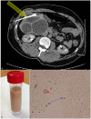
Unusual multiple large abscesses of the liver: interest of the radiological features and the real-time PCR to distinguish between bacterial and amebic etiologies
- PMID: 24548161
- PMCID: PMC4083168
- DOI: 10.1179/2047773213Y.0000000121
Unusual multiple large abscesses of the liver: interest of the radiological features and the real-time PCR to distinguish between bacterial and amebic etiologies
Abstract
We report a rare case of amebiasis generating 19 large liver abscesses. Such a quantity of abscesses is rare, especially when occurring in a young casual traveler without any immunodeficiency disorders. A possible co-infection was excluded. By contrast, the amebic etiology was confirmed by means of serology and real-time PCR.
Keywords: Amebiasis,; Computed tomography,; Entamoeba histolytica,; Multiple abscesses,; PCR.
Figures


References
-
- Stanley SL., Jr Amoebiasis. Lancet. 2003;361(9362):1025–34. - PubMed
-
- Choudhuri G, Rangan M. Amebic infection in humans. Indian J Gastroenterol. 2012;31(4):153–62. - PubMed
-
- Salles JM, Moraes LA, Salles MC. Hepatic amebiasis. Braz J Infect Dis. 2003;7(2):96–110. - PubMed
-
- Wells CD, Arguedas M. Amebic liver abscess. South Med J. 2004;97(7):673–82. - PubMed
-
- Ralls PW, Colletti PM, Quinn MF, Halls J. Sonographic findings in hepatic amebic abscess. Radiology. 1982;145(1):123–6. - PubMed
Publication types
MeSH terms
Substances
LinkOut - more resources
Full Text Sources
Other Literature Sources
